Intervertebral disc disease (IVDD) in dogs is a condition in which one or more of the discs that sit between the bones of the spine compress and/or injure the spinal cord and nerves exiting the spinal cord. The consequences can range from neck or back pain to complete paralysis. Integrative veterinarian Dr. Julie Buzby explains the types, locations, stages, symptoms, diagnosis, treatment, and prognosis for IVDD.
The spinal cord helps control your dog’s every step, stretch, wag, and jump, actions that are easy to take for granted. Unfortunately, though, spinal cord injury or compression, as occurs in intervertebral disc disease (IVDD), can cause those everyday abilities to change in an instant.
What is IVDD in dogs?
Intervertebral disc disease (IVDD) is a common cause of neck and back pain, weakness, and sometimes even paralysis in dogs. It occurs when the discs between the vertebrae intrude into the spinal canal, leading to injury and/or compression of the spinal cord and associated nerves. IVDD most commonly occurs in the vertebrae of the neck (cervical IVDD) or the mid-back (thoracolumbar IVDD). But it can affect dogs anywhere along the length of the spinal cord.
Spinal cord anatomy
The spinal cord, which sends messages between the brain and body, sits within the spinal canal, a channel that runs through the upper portion of the vertebrae. Below the spinal cord and between the solid portions of the vertebrae lie the intervertebral discs. They have a fibrous outer layer (annulus fibrosis) and a flexible, gelatinous inner layer (nucleus pulposus). This arrangement helps the discs act as shock absorbers.
What causes IVDD in dogs?
IVDD is caused by an intervertebral disc rupturing or bulging upward into the vertebral canal. The vertebral canal can’t increase in diameter to make room for the disc material, and sometimes the disc material can take up more than 50% of the diameter of the spinal canal.
This can lead to significant spinal cord compression. Plus, in several forms of IVDD, the disc material hits the spinal cord with significant force. The end result is bruising and damage to the spinal cord, which, like compression, happens from the outside of the spinal cord.
The specific arrangement of the nerve fibers in the spinal cord helps predict the symptoms a dog will show with different levels of compression and bruising.
- The fibers in the spinal cord that send information about proprioception (position of the feet in space) are fairly superficial.
- Buried a bit deeper is an area of motor fibers that carries “movement” signals to the muscles.
- The pathways for deep pain sensation are deeper still.
This means that a dog with a milder case of IVDD may just lose some perception of where his or her feet are. If there is more significant compression, the dog also becomes weak or paralyzed. Finally, with severe compression, the dog loses deep pain sensation, too.
What are the IVDD locations in dogs?
IVDD can happen anywhere along the dog’s spine. However, approximately 65% of cases of IVDD are confined to the thoracolumbar (mid-back, where the chest and abdomen meet) region. Roughly 18% of IVDD cases only affect the neck. The remainder of cases involve a combination of locations or occur closer to the tail in the lumbosacral vertebrae.
Next, let’s take a look at each location.
Thoracolumbar IVDD
Approximately 77-80% of cases of sudden-onset thoracolumbar IVDD occur between the 11th thoracic vertebra (T11) and the first lumbar vertebra (L1). This region includes the T11-T12, T12-T13, or T13-L1 disc spaces.
Since the disc herniation occurs after the nerves heading to the front legs exit the spinal cord, symptoms only affect the rear legs, bladder, and/or bowels.
Cervical IVDD
The most common location for cervical (neck) IVDD is between the second (C2) and third (C3) cervical vertebrae. Interestingly, Dachshunds, Beagles, or Poodles make up about 80% of cervical IVDD cases.
Generally speaking, dogs with cervical IVDD are more likely to have severe neck pain than weakness or paralysis. You might also notice your dog limping on a front leg if the disc is pressing on a nerve root. In the event the dog does experience weakness, it may occur in all four limbs (tetraparesis). Or it might be more pronounced on one side of the body (hemiparesis).
Lumbosacral IVDD
While uncommon, lumbosacral IVDD, or cauda equina syndrome, can also develop in dogs, affecting the lower back and tail. The most common site for this lesion is at the junction of the lumbar (L7) and sacral (S1) vertebrae.
Affected dogs may experience pain near the site of the disc herniation and hind leg weakness or paralysis. Some also have a limp tail, paresthesia (a pins-and-needles feeling that may lead to self-mutilation), and urinary and fecal incontinence.
What are the types of IVDD in dogs?
What happens within the disc itself, how quickly the changes happen, and the consequences of IVDD can vary. Thus, veterinarians classify IVDD into four types: Hansen Type I, Hansen Type II, Acute Non-Compressive Nucleus Pulposus Extrusion (ANNPE), and Hydrated Nucleus Pulposus Extrusion (HNPE).
To better understand these classifications, let’s distinguish important factors for each type of IVDD.
Hansen Type I IVDD (disc extrusion)
Hansen Type I is the most common type of IVDD in small-breed dogs. It is typically acute in onset, leading to a rapid transition from normal to painful, with varying degrees of mobility.
In IVDD Type I in dogs, the inner nucleus pulposus becomes mineralized and loses its ability to serve as a shock absorber. Abrupt movements cause the mineralized bits of disc material to rupture through the outer annulus fibrosis.
If the mineralized disc material herniates up toward the spine (dorsally), it causes compression and bruising of the spinal cord.
When the disc material shoots more sideways (laterally), the spinal nerves get pinched at the point where they branch from the spinal cord.
Typically, type I IVDD occurs in the neck or thoracolumbar region. Chondrodystrophic breeds (dogs with long bodies and short legs) make up much of the list of predisposed breeds for Type I IVDD, which includes:
- Basset Hound*
- Beagle*
- Bichon Frise*
- Cavalier King Charles Spaniel
- Cocker Spaniel (American and English)*
- Corgi (Cardigan and Pembroke Welsh)
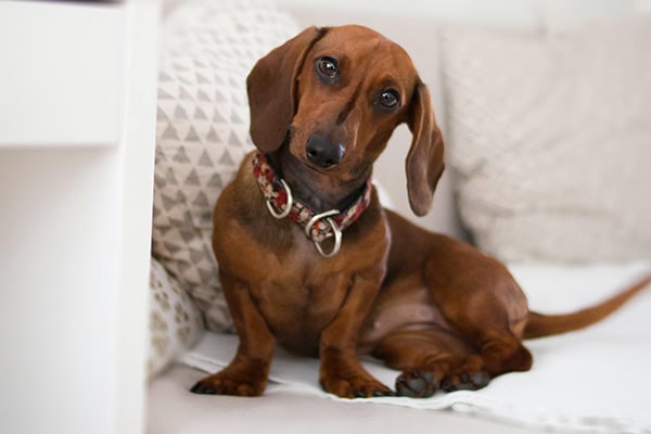
- Dachshund*
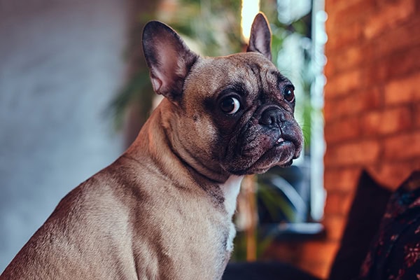
- French Bulldog*
- Lhasa Apso
- Pekingese
- Poodle
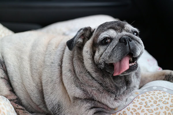
- Pug*
- Shih Tzu*
*These breeds have a genetic marker (an FGF4 retrogene on Chromosome 12). This puts them at the highest risk for developing IVDD. More on this shortly.
Hansen Type II IVDD (disc protrusion)
In contrast to the rapid-onset nature of IVDD Type I, Type II IVDD typically affects dogs more gradually. It is caused by the annulus fibrosus degenerating over time. Eventually, the annulus pushes upward, causing chronic pain and compression of the spinal cord.
Most of the time, IVDD Type II in dogs takes months, if not years, to develop.
Older large breed dogs tend to be overrepresented, with predisposed breeds for Type II IVDD including:
- Bernese Mountain Dog
- Boxer
- Dalmatian
- Doberman Pinscher
- German Shepherd Dog (Considered by many veterinarians to be the poster child for IVDD Type II)
- Labrador Retriever
Generally, IVDD Type II occurs in the thoracolumbar or lumbosacral (pelvic) region. Plus, the dog may have arthritis within the vertebral column.
Acute Non-Compressive Nucleus Pulposus Extrusion (ANNPE)
Acute non-compressive nucleus pulposus extrusion (ANNPE) typically occurs in young active dogs and involves the rupture of a normal disc. A small amount of nucleus pulposus bursts into the vertebral canal under high pressure and at high speed.
Because the extrusion occurs with such force, the spinal cord often suffers a quick, concussive blow and a bruising injury rather than ongoing compression. Think of it like being punched really hard during a boxing match.
Typically, the force of the extrusion may briefly compress the spinal cord. Then the small amount of disc material disperses within the vertebral canal. However, in some cases, the disc material can penetrate the protective lining of the spinal cord (dura mater) or even the spinal cord itself.
Hydrated Nucleus Pulposus Extrusion (HNPE)
Hydrated Nucleus Pulposus Extrusion (HNPE) also involves soft, gelatinous disc material squeezing out of the disc rather than the hard, mineralized material seen in classic IVDD type I.
Unlike in ANNPE where the disc material hits and runs, the soft jelly-like disc material in HNPE acts like a water balloon pressing against the spinal cord. This can cause significant compression.
What are the risk factors for IVDD in dogs?
IVDD has the potential to affect dogs of any size, breed, or age. However, there are a few risk factors that can play a role in making certain dogs more susceptible than others. These risk factors include genetics, body conformation, weight, activity level, trauma history, spay/neuter status, and even diet.
Let’s take a look at each of the risk factors for IVDD.
Genetics
Veterinary researchers have discovered that a genetic mutation, the FGF4 retrogene on chromosome 18, is responsible for creating a short-legged body shape (chondrodysplasia or CDPA).
They also found a similar genetic mutation, called CDDY, that involves the insertion of an FGF4 retrogene on Chromosome 12.
This mutation is linked to early breakdown of the spinal discs in chondrodystrophic dogs. It increases the risk of those discs herniating (rupturing or bulging), which can lead to IVDD.
The FGF4 retrogene on chromosome 12 is considered a dominant gene. This means dogs only need one copy of the gene to have an increased risk of developing IVDD. However, dogs with two copies may be at an even higher risk.
If you are interested in exploring this further, the UC Davis Veterinary Genetics Lab provides testing for the CDPA and CDDY mutations in dogs. Plus, the testing website includes a handy table of the frequency with which CDDY occurs in 64 dog breeds.
Body conformation
As just discussed, chondrodystrophic “low rider” breeds like Dachshunds and Corgis are generally considered to be at higher risk for IVDD, particularly IVDD Type I.
Weight
Obesity in dogs places more strain on the body over time, particularly the neck, back, and joints. This is especially true in the chondrodystrophic breeds like Dachshunds. But it may also be an issue in larger-breed dogs—German Shepherds, for example—who are predisposed to disc problems.
Additionally, in a study on Dachshunds with IVDD, overweight dogs were overall slower to regain control of their bladder function following decompression surgery than those with an ideal body condition score.
This may indicate that weight plays a role in that portion of recovery. However, in that study, being overweight was not associated with the severity of the IVDD, days in the hospital, or days to walk without help.
Activity level
Dogs that compete in frisbee, agility, or other activities that require sudden stops and starts, abrupt twists, or high jumps may be at a higher risk of IVDD. These motions increase the chances that the dog will experience compressive and/or twisting forces along his or her spine that could “blow” a disc.
However, it is important to remember that when it comes to IVDD type I, especially, the disc was already abnormal. Even jumping down off the bed could possibly cause enough force to lead to disc extrusion.
Trauma history
The risk of neck, back, and limb injury is higher in dogs who have experienced trauma. This is the case whether the trauma was from a blunt force (like being hit by a car) or from a fall from a height (down a staircase or off an elevated deck or porch).
Spay or neuter status
Some studies suggest that spayed females have a higher risk of developing IVDD than unspayed females. This may be due in part to weight gain after surgery or the loss of hormones, like estrogen, that play a role in supporting the body’s muscles, joints, and spinal tissues.
In male dogs, the research is less clear. However, because neutering can also lead to weight gain and changes in hormone levels, some researchers believe similar concerns may apply to neutered males as well.
Diet (home-cooked vs commercial)
Home-cooked diets can be fed safely when formulated with the help of a veterinary nutritionist. However, DIY versions often run the risk of not being fully balanced. Over time, those ingredient gaps can matter, especially for dogs with conditions like IVDD, where the body benefits from optimal nutrition to support muscle strength, nerve health, and recovery.
Thus, feeding your dog a commercial diet that is well balanced and nutritionally complete may help decrease the risk of IVDD.
What are the IVDD symptoms in dogs?
IVDD symptoms in dogs can vary greatly depending on the severity and location of the disc herniation and the type of IVDD.
Early signs of IVDD in dogs
Because IVDD type I, ANNPE, and HNPE all happen suddenly, there may or may not be any early warning signs. Your dog can seem totally normal, then rapidly become painful and/or weak. (See below for more information on what pain and weakness look like in dogs with IVDD). Because a dog can go from mildly painful or weak to paralyzed in a matter of hours in some cases, it is important to recognize the signs and seek care immediately.
With type II IVDD being slow and progressive, there may be more opportunity to catch it early. You may notice that your older dog seems to be having just a little more trouble getting up, doesn’t want to walk quite as far, or has more of an unsteady gait. The challenging thing here is that those signs of type II IVDD also accompany osteoarthritis in dogs and a host of other orthopedic conditions.
Common signs of IVDD in dogs
Dogs with IVDD may have some of the following symptoms in varying degrees: pain, changes in posture, a stiff-legged gait, balance problems, knuckling, and weakness, just to name a few. Plus, the signs may show up suddenly or happen gradually.
Let’s take a look at each of the common signs of IVDD in dogs.
Pain
Dogs with IVDD at any point along the spine will experience varying degrees of pain, from mild to severe. This is due to the compression and injury of the spinal cord, spinal nerves, and the highly innervated dorsal longitudinal ligament that runs along the floor of the spinal canal.
Dogs with IVDD may yelp when petted, picked up, or when moving their head or spine. Or you may notice more non-specific signs your dog is in pain.
Changes in posture
Depending on where the pain is, the dog may assume different standing positions:
- Hunched or curved back: Many dogs with thoracolumbar IVDD will try to relieve their discomfort by bowing up their back to alleviate pressure at the site of the spinal compression. These dogs often resemble inchworms with a pronounced curve along the spine.
- Reluctance to move the head and neck: Dogs with cervical IVDD often walk with their necks stiff and straight. Many are unwilling or unable to turn their heads to the side, look up, or lower their heads to reach their food and water bowls.
Stiff-legged gait
A stiff, stilted gait may be noticeable in dogs with an IVDD lesion in the neck or back. This change in gait and mobility is due to pain from compression of the spinal cord or spinal nerves.
Looking wobbly when walking or standing
IVDD of the neck or back may cause the dog to be wobbly and off balance (ataxic). Compression and/or bruising of the spinal cord disrupt the signals that normally travel between the brain and the body via the spinal cord, making it hard for the dog to balance and walk.
- Dogs with IVDD in the back may have a drunken stagger or an exaggerated swishing gait in the back legs as the front legs work to support the body.
- Dogs with mild cervical IVDD may walk with a wide-based, “Bambi on ice” gait if all four limbs are affected.
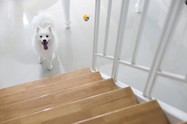
Refusal to jump or navigate stairs
Dogs with cervical IVDD may refuse to walk down stairs or jump off furniture. This is likely due to the compressive forces they experience when their front feet impact the ground.
By contrast, dogs with thoracolumbar IVDD may refuse to climb up stairs or jump onto furniture. The force required to propel them up and/or forward can cause back pain, so they want to avoid it.
Paws knuckling
Knuckling in dogs occurs when they lose the ability to reliably place their foot on the ground in its normal anatomic position. Instead, they will walk on the tops of their toes or drag the toes along the ground. This is one of the earliest signs of IVDD in dogs.
As you learned, the nerves carrying information about proprioception (position of the feet in space) are more superficial in the spinal cord than those for movement and deep pain. Thus, it doesn’t take as much compression or damage to affect them.
Weakness
Moderate IVDD also causes weakness in the limbs. The dog may struggle to get up, stand up and then collapse again, or be unable to stand or walk without assistance.
- Dogs with thoracolumbar IVDD only have weakness in the rear legs (paraparesis)
- Cervical IVDD sometimes causes weakness in all four limbs (tetraparesis) or on one half of the body (hemiparesis)
Paralysis
Paralyzed dogs are unable to voluntarily move the affected limbs at all. THIS IS AN EMERGENCY.
Difficulty urinating
Dogs who are painful or weak but still able to walk may struggle to maintain the posture needed to urinate. This can lead to the bladder being fuller than usual. They are still able to initiate urination, though.
On the other hand, dogs with more severe cervical or thoracolumbar IVDD may lose the ability to voluntarily urinate. Their bladder continues to fill with urine, and the spinal cord can’t send the message to empty it. This can lead to a very full and firm bladder that is difficult to express manually, called an “upper motor neuron bladder.”
Finally, dogs with lumbosacral IVDD may have urinary incontinence in dogs due to a “lower motor neuron bladder.” Their bladder will be large and flaccid, and they may frequently drip urine.
Fecal incontinence
Dogs with mild IVDD symptoms may struggle to have a bowel movement because they are unable to stand comfortably to defecate.
By contrast, fecal incontinence in dogs may occur with severe IVDD symptoms when the nerves that control the anal sphincter become compressed or damaged. The end result is the passive expulsion of feces that the dog cannot control.
Behavior changes
Dogs in pain from IVDD may growl, snap, or bite out of fear or as a protective mechanism. Thus, it is important to be careful when touching, moving, or picking them up.
Loss of appetite
This could be a genuine decrease in appetite due to pain. Or the dog might simply be too painful to walk or move his or her head to reach the food bowl.
When do I need to take my dog to the vet?
If your dog has suddenly become paralyzed, make an emergency vet visit immediately. The sooner your dog can undergo surgery to relieve the spinal cord compression, the better the prognosis generally is.
Dogs who have rapidly become weak and painful also need prompt veterinary attention.
Slowly progressing weakness (over weeks to months) isn’t generally an emergency, but you should schedule an appointment with your vet.
Prior to getting to the vet, try to keep your dog as still and calm as possible. Jumping, running, and playing can worsen the IVDD and your dog’s overall prognosis.
HELPFUL TIP: If your dog is having trouble walking or cannot walk, most clinics have a wheeled gurney or a portable stretcher they can use to safely transport your dog from your vehicle into the clinic. You can give your vet a call as you arrive, so they can be prepared. They may also recommend placing a soft muzzle on your dog during transport for everyone’s safety. Even the nicest dog may bite when experiencing significant pain.
How is IVDD diagnosed in dogs?
The vet will base the diagnosis of IVDD on the dog’s breed, history, symptoms, neurologic exam findings, and imaging results.
Gathering a history
Once in the exam room, the veterinary team will collect a detailed history from you. This includes asking about rigorous activity, falls, or other types of trauma that may have contributed to the symptoms.
Performing the physical and neurologic exam
After collecting the history, the veterinarian will perform a thorough physical exam on your dog. Then, once the initial examination is complete, the next step is a neurological examination.
Some potential tests your vet may perform include:
- Palpating the spine and neck to check for pain, a common finding in IVDD
- Conscious proprioception in dogs (When the vet turns the toes under, dogs with IVDD may leave the foot knuckled or be slow to return the foot to a normal position.)
- Reflex testing (Responses may be absent, delayed, normal, or exaggerated.)
- Cutaneous trunci reflex (When the vet pinches the skin by each vertebra of the mid and lower back, it should twitch. If it doesn’t, the spinal cord lesion is usually about 1-2 vertebrae toward the head from where the reflex cuts off.)
- Evaluating tail movement and anal tone (Some dogs with IVDD lose these functions.)
- Assessing the deep pain response (When the vet pinches a toe, does the dog turn or yelp, showing he or she felt the pain? Just pulling away is a reflex and does not indicate deep pain is present.)
These tests help confirm there is a neurological problem and localize where the spinal cord seems to be damaged. Plus some tests, like deep pain, provide information about prognosis.
X-rays
X-rays are a great way to screen for other problems with the spine, such as broken or dislocated bones or bony bridging between the vertebrae (spondylosis in dogs). The vet can also look for mineralization of discs between the vertebrae, the collapse of normal disc spaces, narrowing of the intervertebral foramen (“horse head” shaped spaces between vertebrae where nerves exit the spinal cord), or other changes that point to IVDD.
Since IVDD causes pain at every stage, your vet will likely recommend either a light plane of sedation or general anesthesia for the X-rays. Sedation allows the veterinary team to position your dog for imaging without causing him or her pain or having your dog struggle while in an awkward position. And it helps the vet get good quality diagnostic images, too.
It is important to note that while X-rays can point to IVDD, they are not sufficient for surgical planning. A dog needs advanced imaging performed by a veterinary specialist before IVDD surgery.
Consultation with a neurologist and advanced imaging
If surgery is a consideration or your dog has a complex case, your vet may refer your dog to a veterinary neurologist. This specialist can review the test results and notes from your vet and decide which additional imaging will be most appropriate for your dog.
Specialized imaging tests include:
- Myelography: This study involves injecting an iodine-based dye around the spinal cord. Then, the veterinary team takes an X-ray to look for narrowing of the dye column at a point of compression.
- MRI: An MRI can identify disc degeneration earlier than a CT scan and allows the vet to visualize the spinal cord, bone, and soft tissues. Plus, it can provide prognostic information.
- CT scan: This scan allows the vet to see cross-sectional images of the spine at the level of compression.
NOTE: Depending on the imaging results, these cases often go directly to surgery. Time is of the essence in relieving compression on the spinal cord and nerves.
Stages of IVDD in dogs
Based on the diagnostic information, the vet will determine which stage of IVDD your dog has. (The term “grade” can be used interchangeably with “stage” here.)
Stage 1: Mild pain
Typically, Stage 1 is the mildest episode of IVDD that a dog experiences. Affected dogs may feel mild neck and/or back pain that resolves after a few days. Stage 1 IVDD is a non-surgical diagnosis. Supportive care, like exercise restriction and NSAIDs, usually leads to complete recovery in 70-100% of cases within a few days.
Stage 2: Moderate to severe pain, mild weakness, or wobbly gait
Dogs with Stage 2 IVDD may have moderate to severe pain in the neck or back with mild weakness or a wobbly gait. They are generally still able to stand and walk on their own. Stage 2 is usually non-surgical unless the dog’s symptoms progress in spite of conservative treatment. Approximately 55-100% of Stage 2 IVDD dogs will recover to walk normally or close to normal with rest and conservative care.
Stage 3: Significant pain and weakness
For dogs experiencing Stage 3 IVDD, the treatment protocol often shifts from non-surgical to surgical. Symptoms at this stage include significant pain and weakness (paresis), meaning the dog cannot stand or walk without help. With surgery, roughly 80-90% of Stage 3 IVDD dogs will be able to walk again, although they may never return to a completely normal gait. However, this range drops to 55-80% in dogs who do not undergo surgery.
Stage 4: Paralysis with deep pain present (Emergency situation)
The hallmarks of stage 4 IVDD are paraplegia (paralysis in the rear limbs) or tetraplegia (paralysis in all four limbs), depending on the level at which spinal compression occurs. Stage 4 dogs experience loss of bladder function, but still have deep pain present. This stage is considered an EMERGENCY.
In many cases, dogs with Stage 4 IVDD will be moved directly from imaging under anesthesia to surgery in an effort to save the spinal cord from permanent damage. Recovery from Stage 4 IVDD can take up to two months following surgery. Upwards of 80-90% of dogs will regain the ability to walk. Like Stage 3, though, their gait may never be completely normal.
Stage 5: Paralysis with NO deep pain (Emergency situation)
The most serious type of IVDD, Stage 5, is characterized by paraplegia or tetraplegia and loss of bladder function with NO deep pain present. Accordingly, Stage 5 IVDD is an absolute EMERGENCY, necessitating immediate surgery.
Unfortunately, for dogs with Stage 5 IVDD, only about 50% of these dogs will walk again, even with surgery. In these cases, recovery time ranges anywhere from roughly two to nine months.
What is the treatment for IVDD in dogs?
Treatment for IVDD in dogs often varies based on the severity and duration of the symptoms. Dogs with Stage 1 or 2 IVDD are usually able to be treated conservatively at home. However, surgery provides the best outcome for most dogs with Stages 3 and above (or dogs who were Stage 2 but continue to progress).
Medical treatment of IVDD
Conservative treatment for IVDD in dogs is typically prescribed if the dog is able to walk, has pain perception in his or her feet, and has bladder and bowel control. If the dog has more severe IVDD but surgery isn’t an option due to finances or the dog’s health concerns, the vet may also try medical management.
However, it is important to understand that higher-grade IVDD isn’t the sort of condition where you can try conservative treatment and then move on to surgery if it doesn’t work. If you are going to do IVDD surgery, it needs to be done ASAP.
It is imperative to follow your vet’s instructions closely to ensure your dog’s best chance of recovery.
Strict crate rest
Dog crate rest is one of the most important (and challenging) parts of non-surgical treatment of IVDD. Ideally, you would leash-walk or carry your dog outside to potty, then bring him or her directly back to the crate. No sniffing around the yard, hanging out on the couch with you, playing with other pets, etc.
The minimum period for crate restriction is typically four to six weeks. But it is often closer to eight weeks. This may seem like a long time at the outset, but it is vital for your dog’s recovery.
Note: Dogs with cervical IVDD should be carried, not led outside on a leash. This helps avoid causing pain or additional injury if the leash pulls taut around the neck.
You should NOT assume your dog will self-isolate or curb his or her activity due to pain. Instead, stick to the crate rest plan laid out by your vet. Not doing so can worsen your dog’s IVDD to the point that he or she needs surgery.
Medication for IVDD in dogs
Your vet may also prescribe the following medications:
- Steroids or NSAIDs: Your vet may prescribe steroids (prednisone for dogs) OR non-steroidal anti-inflammatory drugs (NSAIDs) such as carprofen for dogs. These medications help decrease inflammation around the affected disc.
- Dogs should NOT take steroids and NSAIDs together due to the risk of gastrointestinal upset and the possibility of causing stomach ulcers in dogs.
- Antacids: The vet will often also prescribe an antacid like omeprazole for dogs or Pepcid for dogs. This can help protect the stomach lining while the dog is on steroids or NSAIDs.
- Muscle relaxers: Medications like Robaxin® (methocarbamol for dogs) and diazepam are helpful for relieving painful muscle spasms that often develop around the site of the disc herniation. (They don’t directly address pain, though.)
- Gabapentin: Because it is good for neuropathic pain, vets often prescribe gabapentin for dogs to address pinched spinal nerves. It tends to be part of the multimodal pain management plan in conjunction with steroids or NSAIDs.
Integrative treatments
Integrative medicine approaches such as acupuncture for dogs, laser therapy for dogs, rehabilitation therapy, and PEMF for dogs also make great additions to the treatment plan. They are not, however, substitutes for traditional medical treatment of IVDD, and not all options are right for all dogs.
Your vet can help you determine which approaches your dog may benefit from. Or he or she may refer you to another veterinarian with more experience in integrative medicine.
Note: Some of these therapies are also beneficial for dogs after IVDD surgery. The neurologist will advise you on the options.
Surgical treatment for IVDD
For the best chances of recovery, dogs with IVDD type I who are significantly painful and weak or paralyzed (stages 3-5) need immediate surgery to relieve the spinal cord compression. Additionally, surgery may be recommended for dogs with Type I Stage 2 IVDD who are not improving with medical management and for some cases of IVDD Type II. ANNPE is non-surgical, and HNPE is sometimes treated with surgery.
The neurologist will determine which surgical procedure is best suited for your dog’s particular situation. Some possible types of IVDD surgery for dogs include:
- Hemilaminectomy: Creating a “window” on one side of the vertebral canal to relieve compression
- Dorsal laminectomy: Removing the whole “roof” of the vertebrae
- Pediculectomy/mini-laminectomy: Similar to the hemilaminectomy, but removes less bone, so it is less invasive
- Ventral slot: Addressing a herniated disc in the neck from below rather than above
- Prophylactic fenestration: Making openings in the surrounding discs to decrease the chances they will herniate into the spinal canal
Post-op care from IVDD surgery
When your dog comes home from surgery, the veterinary team will discuss the specifics of caring for him or her. Your dog may or may not be able to walk yet.
For dogs who need some mobility help, the Help ‘Em Up® Mobility Harness makes it easier to support your dog. Dr. Buzby’s ToeGrips® dog nail grips are also great for improving your dog’s traction and sense of where his or her feet are.
If your dog can’t get up on his or her own, it will also be important to provide soft bedding and reposition your dog frequently. This can help prevent pressure sores on dogs.
Additionally, you may need to express your dog’s bladder (your vet will teach you how) and keep a close eye on his or her ability to urinate. Plus, some dogs require frequent cleaning due to fecal or urinary incontinence.
Are there home remedies for IVDD in dogs?
There are no effective home remedies for IVDD. Nothing takes the place of a veterinary evaluation, thorough diagnostics, and a comprehensive management plan. Waiting to seek veterinary care for dogs who are suddenly weak, painful, or unable to walk can lead to devastating consequences and a much poorer prognosis for recovery.
What is the prognosis for IVDD in dogs?
The long-term prognosis for dogs with IVDD depends on many factors, like the severity of the episode, breed and age of the dog, other medical issues the dog is experiencing, and the length of time between the first onset of symptoms and treatment.
As a general rule, the longer the neurologic symptoms have been present, the poorer the prognosis.
Overall, the prognosis is good for dogs with mild IVDD (Stages 1 and 2). With conservative management and careful adherence to the plan, these dogs have a good chance of recovering well and walking normally.
However, dogs have an increasingly poor prognosis as the stage increases. If a dog cannot walk but has deep pain in at least one leg, the surgical success rate is 83-93% for recovery.
If a dog cannot walk and has no deep pain, but has been symptomatic for less than 48 hours, the surgical success rate is roughly 50-60%. Unfortunately, if a dog’s symptoms have been present longer than 48 hours, the surgical success rate is dramatically lower. At this point, surgery may not be worth pursuing.
It is also important to remember that while IVDD can be treated, it isn’t necessarily “cured.” Dogs who have had one disc rupture are more susceptible to a subsequent episode. Usually, it is the disc on either side of the first one that ruptured. In these cases, prophylactic fenestration may be particularly helpful in minimizing that risk. Many veterinary surgeons perform that procedure at the same time as the decompression surgery.
How do you prevent IVDD in dogs?
Unfortunately, there’s no guaranteed way to prevent your dog from developing IVDD. However, there are steps you can take to help reduce the risk and support your dog’s spinal health.
- Pursue genetic testing for IVDD to help you better understand your dog’s risk level.
- Find your dog’s body condition score (BCS) and keep him or her at an ideal weight.
- Engage your dog in regular low-impact exercise to build and maintain muscle mass.
- Select activities or games that minimize the amount of jumping and twisting your dog does.
- Use stairs or ramps (like Doggo Ramps™) to minimize jumping on and off the furniture.
Act fast if you see signs of IVDD
If you notice signs of pain, weakness, wobbliness, or difficulty walking in your dear dog, it’s important to seek veterinary care as soon as possible. Prompt diagnosis and treatment can make a huge difference in your dog’s comfort, recovery, and long-term mobility. In many cases, timely care improves the chances of a full recovery. And it helps reduce the risk of permanent pain or neurologic damage.
Has your dog ever been diagnosed with IVDD?
Please share your experiences below.