Histiocytoma vs. mast cell tumor—what’s the difference between these two similar-looking skin tumors in dogs? Integrative veterinarian Dr. Julie Buzby compares and contrasts the appearance, location, treatment, and prognosis for histiocytomas and mast cell tumors. Discover why it is critical to distinguish between the two—for the sake of your dog’s health.
Lumps and bumps can pop up on our dogs and cats at any time. And they run the gamut from benign skin tags to serious cancers. But when it comes to skin masses that are very common in dogs and look similar, like cutaneous histiocytomas and mast cell tumors, how do you know which bump is which? And does it matter?
As it turns out, it is important to know if your dog has a histiocytoma vs. mast cell tumor. The treatment and outlook are very different for these two look-alike tumors.
What is a histiocytoma?
Histiocytomas are a common type of skin tumor in dogs, and they’re almost always benign. As the name would imply, they occur due to abnormal multiplication of histiocytes (i.e. histiocytic cells). These cells are a precursor to macrophages, Langerhans cells, and other cells that play an important role in the immune system. Typically, the cells that form the histiocytoma are Langerhans cells that migrate from the middle skin layer to the surface of the skin and make a mass. However, no one knows why cutaneous histiocytomas in dogs form.
What is a mast cell tumor?
Mast cell tumors are the most common type of skin cancer in dogs. They arise from the uncontrolled replication of abnormal mast cells. Mast cells are also an important part of the immune system. They contain granules filled with substances such as histamine, growth factors, and heparin. When mast cells are part of a tumor, they may release these chemicals in an uncontrolled manner. This can lead to blood-clotting problems, GI ulcers, and anaphylaxis (a severe type of allergic reaction).
Histiocytoma vs. mast cell tumor: Appearance
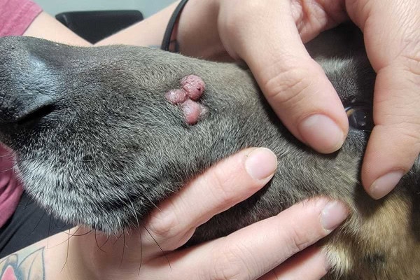
Cutaneous histiocytomas tend to be round, firm, raised skin masses that can have some or no hair on them. They can be pink or red, and have distinct borders. Typically, histiocytomas are no larger than two to four centimeters wide. Since they sort of look like a pink button on your dog’s skin, histiocytomas have earned the name “button tumors.”
The picture below illustrates the button-shaped appearance of a histiocytoma.
On the other hand, mast cell tumors can be subcutaneous (i.e. under the skin) or cutaneous (i.e. on the skin). This is unlike histiocytomas, which only occur on the skin, not under it. Mast cells tumors on the skin have many of the same characteristics as histiocytomas. They tend to be pink or red hairless tumors with distinct borders.
However, mast cell tumors in dogs can also be other colors like white or tan. In fact, they can look like almost any other kind of skin bump, including lipomas in dogs. This is part of what makes mast cell tumors so dangerous.
Where do histiocytomas tend to occur?
Histiocytomas can occur anywhere on the dog’s body. But one study demonstrated that 29% of histiocytomas occurred on the head, and almost half of those were on the ears. The most common locations for dog histiocytomas to form are:
- On the muzzle, lips, or bridge of the nose
- On a dog’s paw or leg
- On the ear flaps (i.e. pinnae)
- On the chest
- Anywhere on the neck
- Around the eyes and on the eyelids
Where do mast cell tumors arise most often?
Mast cell tumors can also occur anywhere on the body. Unlike histiocytomas, there is no tendency to form in any particular location. They can be on the dog’s head, neck, chest, trunk, belly, legs, or tail.
Are there breed or age predispositions for each tumor type?
While any dog of any age could technically develop a histiocytoma or mast cell tumor, certain dogs do tend to be prone to each tumor type.
Histiocytoma
Overall, histiocytomas tend to occur in young dogs (less than three years of age). And they are the most common skin tumor of dogs under a year of age. However, middle-aged and older dogs do sometimes develop histiocytomas too.
Histiocytomas are more likely to form in purebred dogs such as Scottish Terriers, Bulldogs, Great Danes, and Cocker Spaniels. Flat-coated Retrievers are four times more likely to form histiocytomas when compared to other breeds. And the Chinese Shar Pei is the most likely breed to develop multiple histiocytomas.
Mast cell tumor
In contrast to histiocytomas, middle-aged or older dogs are more commonly affected by mast cell tumors. But younger dogs are not immune. The following dog breeds have a higher risk for developing mast cell tumors:
- Beagles
- Boston Terriers
- Boxers
- Chinese Shar Peis
- Cocker Spaniels
- English Bulldogs
- Golden Retrievers
- Labrador Retrievers
- Weimaraners
Histiocytoma vs. mast cell tumor: How does each behave in the body?
These two tumor types may act very differently in the body. This is one of the reasons it is so important to know which tumor your dog is dealing with.
Histiocytomas—usually benign but can be cancerous in rare cases
Thankfully, almost all histiocytomas are benign and are unlikely to spread to the internal organs. Histiocytomas usually grow rapidly for the first few weeks but then resolve on their own after about one to three months. They generally aren’t itchy or painful, although histiocytomas that develop on the eyelid may cause slight discomfort.
Typically, the dog will develop one histiocytoma at a time. But sometimes dogs can develop cutaneous Langerhans cell histiocytosis (LCH). In this condition, the dog may have multiple histiocytomas at once and the skin is ulcerated and irritated. LCH may also impact the mouth and the junction between haired skin and mucous membranes, which is different from typical histiocytomas.
In very rare circumstances, a malignant type of histiocytoma can form. Also known as systemic histiocytosis or histiocytic sarcoma complex, malignant histiocytosis is a disorder found primarily in Bernese Mountain Dogs. But it has also occurred in some Golden Retrievers, Flat-coated Retrievers, and Rottweilers.
In malignant histiocytosis, multiple histiocytomas may form on the skin, subcutaneous tissue, or joints. But they also occur in major internal organs like the lungs, liver, and spleen, as well as regional lymph nodes and the nervous system. Sadly, malignant histiocytosis is a fatal disease with a very short survival time.
Mast cell tumors—cancerous
Unfortunately, mast cell tumors in dogs are cancerous. And they can spread to the lymph nodes and bone marrow. Mast cell tumors may also affect the spleen and liver, but rarely spread to the lungs. Additionally, they may result in systemic clinical signs such as vomiting, diarrhea, fever, swollen limbs, and collapse.
How do you know if your dog has a histiocytoma or a mast cell tumor?
With most histiocytomas being no big deal and mast cell tumors being very much a big deal, it is important to be able to know which tumor your dog has. As you have learned, both can look similar, so there really isn’t a good way to visually distinguish between the two. Plus, mast cell tumors have earned the moniker “Great Pretender” because they can look like anything—not just like a histiocytoma.
Sure, histiocytomas usually affect young dogs, and they can resolve on their own after a few months. However, without an official diagnosis, you might wait several months only to find out that the growth you assumed was a histiocytoma didn’t go away because it was actually a mast cell tumor. And now you are months behind in getting your dog the treatment he or she needs.
Knowing all this, the best course of action if you find a mass on your dog’s skin is to make an appointment with your vet. He or she can examine the mass and perform a fine needle aspiration (FNA). This is one of the main ways to identify the various skin tumors dogs may get. Performing an FNA involves inserting a small needle into the mass to withdraw some cells. Then the veterinarian will analyze the cells under the microscope or send the slide to a veterinary clinical pathologist for evaluation.
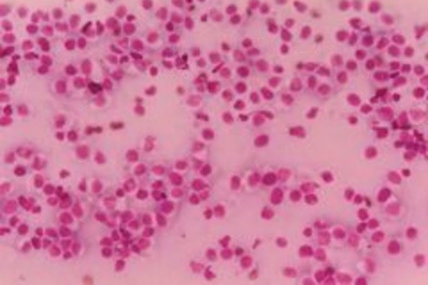
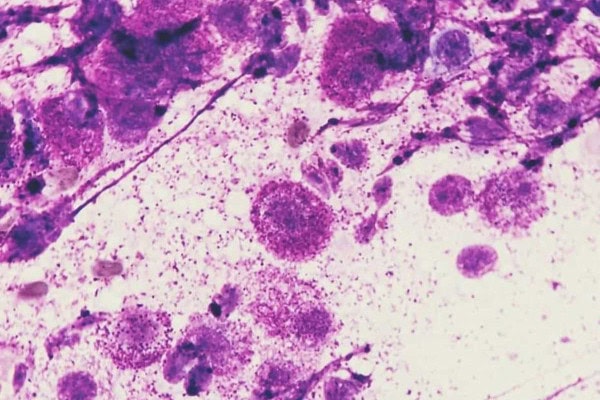
On cytology (i.e. when examined under the microscope), histiocytomas tend to have moderate numbers of histiocytes and some other immune system cells like lymphocytes. And mast cell tumors are composed of mast cells—round cells which usually have prominent granules.
The two images below show the difference between a histiocytoma and degranulated mast cells when viewed under a microscope.
What is the treatment for histiocytomas in dogs?
If the result of the fine needle aspiration shows that the tumor is a histiocytoma, benign neglect (i.e. leaving it alone) is a reasonable treatment option. The histiocytoma will typically start to regress on its own in a few months without the need for any veterinary or home treatment.
However, your veterinarian may recommend cryotherapy (i.e. freezing the tumor) or surgical removal if the histiocytoma doesn’t go away within three months, becomes ulcerated, swollen, infected or starts bleeding, or is painful and growing rapidly.
When a histiocytoma is ulcerated or infected, your veterinarian may also prescribe a course of antibiotics. However, he or she will usually avoid using immunosuppressants because the dog’s own immune response to the histiocytoma is what causes it to go away. However, dogs with multiple histiocytomas may need immunosuppressive or chemotherapeutic therapies.
What is the treatment for mast cell tumor in dogs?
Because they are cancerous, the treatment for mast cell tumors is much more aggressive than for histiocytomas. When possible, the vet will recommend a mast cell tumor be surgically removed. During surgery it is important to take wider margins around the mast cell tumor than are necessary when removing a histiocytoma. This is the case because it is important to ensure all of the mast cell tumor is removed due to its potential to spread.
If the dog has metastatic disease, the vet may recommend radiation and chemotherapy too. Alternatively, an intratumoral injection called Stelfonta may be an option for some mast cell tumors.
Histiocytoma vs mast cell tumor: What is the prognosis?
As you can imagine, dogs with solitary histiocytomas have an excellent prognosis. The tumor almost always resolves within one to three months without any treatment interventions. If surgery is needed, complete removal of the mass is usually curative.
Dogs with Langerhans cell histiocytosis have a more guarded prognosis. Regression of the tumors usually takes about 10 to 12 months. And if LCH spreads to other organs or the skin is severely affected, humane euthanasia may be necessary.
The prognosis for malignant histiocytosis is guarded to grave, unfortunately. It doesn’t respond well to traditional therapies and in a study of histiocytic sarcoma in Bernese Mountain dogs, average survival time was only 49 days after diagnosis.
Thankfully, lower-grade mast cell tumors can behave benignly and surgical removal is often curative. However, higher grade mast cell tumors have a guarded to poor prognosis, depending on their location. And mast cell tumors that form on the muzzle or near a nail bed tend to be associated with a poorer prognosis.
Head to the vet if your dog has a skin tumor
If you only take away one thing from this article, let it be this…
If you find a new lump or bump on your dog, the best thing you can do is to make an appointment with your vet. Without some diagnostic testing (usually a fine needle aspiration) there really isn’t a way to definitively know what type of mass your dog is dealing with.
When it comes to cancerous skin masses, the sooner you find and remove them the better. If you are looking at a mass on your dog’s skin right now and Googling “histiocytoma vs mast cell tumor,” please give your vet a call so you can find the answers that your dog needs.
Has your dog had a histiocytoma or mast cell tumor?
Please comment below.

Our 9 yo Brittany was diagnosed with fine needle biopsy with Mast Cell Carcinoma about 8 months ago. Oncologist said would require deep surgery and then radiation. For connivence we changed Vets and at this time the under skin mass comes up and down and after red surface sore went away, it has never resurfaced. She is active, terrific appetite, keeps up playing and running like our other 2yo Brittany. The current Vet when I asked does not understand her obvious health???? She also said under the microscope it can be vary similar to another non cancer condition????? She is on 50mg Benadryl and and 20mg Famotadine twice daily. She also said a Mast Cell could be disturbed if we did another needle biopsy? We have stopped all travel plans because we dont want to board her with this? That is not the most important but for retired Seniors it is part of the equation. Thanks
Hi Steve,
I am sorry your pup is dealing with this puzzling tumor situation. Yes, some mast cell tumors (MCT) and histiocytomas can seem very similar in the way they appear and behave. It is possible to cause a degranulation episode (the mass releases histamine and many other negative chemicals) by inserting a needle into a MCT which can set off a very bad chain of events. For this reason, anytime we suspect a MCT we premedicate the dog with Benadryl to try and prevent degranulation effects/allergic reaction. I can’t say for sure why the mass has not “resurfaced” and without understanding all the details am not able to offer specific conclusions or recommendations. Unless the cancer has progressed to an end stage, I would not see any reason to avoid boarding or other normal activities. Hoping you can get some clarity and peace about your Brittany’s condition. Wishing you all nothing but the best.
I had to put my 9yr old blue pitbull to sleep in 2024 because he had Hemangiosarcoma and MST. That was the hardest thing I had to do. I adopted a 7 wk old puppy from the shelter a week after. About 2 weeks ago I noticed a bump on the inside of my dog’s toe. Took him to the vet and she gave him steroids and antique because it was infected from him licking it. Vet said it’s more than likely a histiocytoma. She will do another exam on his next check up.
Dear Tricia,
I am sorry for the loss of your senior guy and can only imagine how difficult that must have been. What a blessing your vet is not too concerned about this bump on your new puppy. Hoping it will resolve on its own and you receive good news at the next checkup. Wishing you and your sweet boy all the best for many happy years to come.
Hello
My 4 year old Doberman had two histiocytoma tumors that were recently removed and biopsied. We are awaiting results and a third just popped up on her leg . Im
Curious to understand if both benign and mast cell tumors soread? Thank you
Hi Dee,
I am sorry your girl has been through so much with these frustrating tumors. I am not sure I understand exactly what you are asking. Histiocytomas are always benign, but they are a completely different type of tumor from a mast cell tumor. Mast cell tumors are always malignant but depending on their grade and location, they may be completely removed with surgery or may end up spreading to other areas of the body (metastasize). Not all mast cell tumors are aggressive and there is a better outcome if they are dealt with quickly. Hoping this helps clear things up a bit and praying for favorable results from the biopsies. Happy New Year!
Thank you for your quick response . Apologies about not being clear. We do not yet have an official diagnosis, however I am referring to them as histiocytomas. They look exactly like them and our vet refered to them as such how ever the aspiration reading was suspicious . Here’s what it read : Atypical mesenchymal cell proliferation – most consistent with mesenchymal neoplasia
Comments:
The sample contains an atypical proliferation of mesenchymal cells – aspiration of a mesenchymal neoplasm (eg, soft tissue mesenchymal tumor vs other) is considered most likely. Although very rare histioctoid cells are observed, the cytologic features of this mass are not consistent with a diagnosis of histiocytoma. Surgical excision with histopathology is recommended..
While we await the biopsy results post removal of two, a new third growth has appeared on her leg. I’m curious if it is more common for a histiocytoma or a mast cell to spread on the exterior of the body similar to what we are experiencing? Thank you
Hi Dee,
Ah! I understand now! Thank you for the extra information. Yes, it is much more common for dogs to have several mast cell tumors appear on the skin in different locations on the body. It doesn’t necessarily mean they have metastasized, but rather that the dog’s genetics has a predisposition to making these types of tumors. Here is a link to another article with more information: Mast Cell Tumors in Dogs: Pictures, Prognosis, Diagnosis, Treatment
Hoping the pathology results will offer some insight and help guide you toward the best treatment. Praying for a clear path forward and a positive outcome for your sweet girl. ♥
My dog has a pink hairless lump below his eye but not on the eye it doesn’t seem to be painful for him but it is firm i’m unsure what it could be ?
Hi Zara,
I understand your concern for your pup with the appearance of this lump below his eye. Without being able to examine it myself, it is hard to offer specific conclusions. It would be best to have your vet take a look at the lump and let you know if any testing is required or if removal is recommended. Here is a link to another article: Dog Eyelid Tumors: Types and Treatments
I am not sure if this is what your pup is dealing with, but it may offer some good information and give you an idea of what to discuss with your vet. Hoping for clear answers and an easy solution. Best wishes!
Just came back from the vet. My 9 yo Lab mix has either a histiocytoma or a low grade MCT. We’re going to give it a couple of months to see if it recedes and if it doesn’t resolve, or if it starts to grow or get ugly, we’ll take it off. It just didn’t look like a ‘normal’ growth to me, Glad we went in, I feel much better about her prognosis now.
Hi MaryKate,
I am glad you were able to get this mass evaluated by your vet. Hoping it will resolve itself and surgery won’t be needed. Wishing your sweet girl the best of luck!
my 13year old dog has to lumps on is pads he has bad arthritis but now with there lumps on is pads is walking is bad
Hi Alison,
I am sorry your senior dog is having issues with lumps on his pads. This definitely sounds like something that needs to be evaluated by your vet. Hoping there is an easy way to offer your sweet boy relief and comfort. Wishing you both all the best.
My dog had a squishy cyst that would drain occasionally on top of her head. Now it is bleeding and crusty. She has diarrhea right now and lack of appetite. I wonder if they are related.
Hi Reenie,
I am sorry your pup is having so many issues at once. It sounds like she would benefit from scheduling an appointment with your vet to have things checked out. Hoping for clear answers and an easy solution. Wishing your sweet girl all the best!
my 12 yr old Maltese has a button tumor on her side. This is her 4th one. in different locations on her body. She has been tested with a needle before and it looks to be the same. She has Addison’s Disease, so she sees 2 different vets both confirm it looks to be another tumor. Because of the AD they want to play the wait and see game instead of treatment. however this is the biggest scab I’ve seen its the size of a penny. historically hers start and leave within the same month but this one looks like it will be lasting longer. shes not been prone to itching or licking it but we’ve dressed her in a baby onesie to make sure its left alone.
Hi Emma,
I am sorry your senior girl has recurring issues with these tumors. What a blessing you have such a great veterinary team working to ensure she remains healthy and happy for as long as possible. You are doing a great job managing all of her medical issues and I applaud you for continuing to advocate for her well-being. Wishing you both all the best and keep up the good work!
my bully Pitt is 1 1/2 years old she has a large bump on her lip. it started out small and just grow bigger over the last 2 months. It was red now it’s soft and looks like it is full of liquid or maybe blood.
what is it and how do I get rit of it
Hi Anitra,
I am sorry your young dog has this unknown bump growing on her lip. I understand you are hoping for answers, but without being able to examine your girl I can’t make a diagnosis or offer specific recommendations. I encourage you to make an appointment with your vet and allow them to evaluate this bump. They can let you know what testing will be needed to get a diagnosis and then talk through the different treatment options. Hoping this issue is nothing to worry about and there is an easy solution.
My 4 year old mix (retriever, mini husky, etc.) has a pink lump/bump seemingly out of nowhere.. The vet scheduled him for surgery to remove it. However, it is almost a month away. Why would she not perform an FNA now? I am so heartbroken for my boy. It is all I think about and have weeks still to wait. I feel there is no urgency from our vet and time matters. I would attach a photo but it doesn’t look like I have the option.
Hi Rock,
I am sorry your pup is facing surgery for this unknown lump. Without playing a personal role in your dog’s medical care it is hard to make assumptions as to why certain decisions have been made by your vet. It may be that your vet is fairly confident about the type of mass this is. If surgery is already scheduled, they could be trying to save you money by forgoing the FNA. While an FNA can give you information about what type of mass this MIGHT be, once the mass is removed and sent to the lab for the pathologist to review you will get a report of exactly what kind of mass this is. If having an FNA done now will give you extra peace of mind, don’t hesitate to ask your vet to perform this test. They may not realize it is important to you, so make sure to make your wishes known. Hoping all goes well and praying for a positive outcome for your sweet boy. ♥
My Dutch Shepperd had a mast cell tumor low grade when he was 2 years old. It was removed without further therapy. Now, at the age of 5, a new lump appeared on his paw and we are very worried. Wednesday we have an appointment at the vet and hope it is not a mast cell tumor again. As it is on his paw now, it’s worrysome if it is cancerous as they have to remove it larger than the tumour itself.
Dear Pascale,
I understand your concern for your Shepherd and think it is good you already have an appointment scheduled to get him evaluated by your vet. I too am hoping this lump is nothing to worry about and praying for favorable results. Feel free to leave an update and let us know what you find out. Wishing you both nothing but the best.
I first noticed a sore and a scab that eventually fell off on my 3.5 year old bernese mountain dog about 6-9 months go. Then in July when she was in for her annual checkup there was a marble sized lump on her belly that the vet said she wanted to leave alone unless it changed. Fast forward to last week and it “erupted” into this skin mass that is big and oozy. It has gotten smaller like it did last time (it completely disappeared last time) and so I thought it was the same thing. She is happy, playing, eating, drinking and being her normal fun and playful self so I didn’t think anything of it until she developed a random limp out of nowhere. She is going in for surgery tomorrow morning to have it removed, and the vet said that her breed is prone to hystiosarcomas, but after reading about them, the prognosis is really bad and I’m hoping because it’s a second occurrence of this and it’s been about 6-9 months since the last “flare up” and she’s been fine, that it’s not cancerous.
Hi Lynn,
I am sorry your big girl is dealing with this worrisome mass on her belly. I understand your concern and am glad you are planning to have it removed. How did the surgery go? Did you get results from the pathologist? Hoping all is well and your girl has made a full recovery. Feel free to leave an update if you have a chance. Wishing you both nothing but the best.
my 3 yo female pug suddenly developed a 5mm raised pink colored lesion on her front leg. Our vet thought it looked like a histiocytoma. her recommendation was to monitor for a month then reevaluate. it is shrinking and almost gone after 4 weeks… We are relieved it seems to be behaving like a histiocytoma. My question is do Mast cell tumors ever devolve without treatment. 🤔
Hi Lesli,
I am glad the lesion on your Pug’s leg is shrinking and almost gone. While Mast Cell tumors can increase in size when disturbed or inflamed, they will not completely resolve and go away without treatment or surgical removal. I am hopeful that since this mass is resolving it is probably a histiocytoma and should not cause any further issues. What a happy outcome for your sweet girl! Best wishes to you both!
My whippet had a mast cell tumor removed when she was about 5 years old. It was surgically removed with wide margins and was a low grade tumor. She did not have any follow-up treatment and now she is 14 and has had no recurrence. I have given her turkey tail mushroom powder which may have staved off a recurrence or maybe we were just lucky. Now, at 14, she has a lot of lipomas. One large one was on her chest and was being irritated when she lay down so I had it removed. The others have shrunk somewhat due to a blend of mushroom powder I give her.
Hi Sharon,
This is very interesting! I am so glad your girl has remained free from recurrent mast cell tumors for over 9 years. What a blessing you were able to find a natural way to combat her tumors and she responded so well to the treatment. Thank you for sharing your experience with us. Wishing you continued success and many happy days ahead. ♥
I just took my 3-year-old dog to the vet today. He’s part lab, part puddle, part golden retriever and has a small bump on his ear flap. The vet got a sample with a fine needle and said it was a mast cell tumor. They’ll do surgery in a month to remove the tumor and will send it to pathology. I’m trying to stay positive and hoping he’ll leave many many healthy years after this surgery, but I’m also very sad and very scared 😞
Hi Rosa,
I understand your concern for your young dog. What a blessing your vet was able to get a diagnosis so quickly and has already scheduled surgery to remove the mass. Try not to let the “what ifs” be a heavy burden. Many dogs go on to live normal happy lives after having surgery to remove one of these tumors. Here is a link to another article with more information: Mast Cell Tumors in Dogs: Pictures, Prognosis, Diagnosis, Treatment
Praying for a successful procedure and an easy recovery. Wishing your sweet boy all the best!
My schnauzer was diagnosed with Mast Cell Tumors about 2 weeks ago. Hers are low grade ones. I guess it had good margins but in the last week she has changed her habits. She has started sleeping all the time and she I’d losing weight. She is 7 years old. Could the cancer be inside also. Thank You
Hi Paulette,
I understand your concern for you Schnauzer with these worrisome behavior changes that have recently emerged. I can’t say for sure that these issues are cancer related but think they should be investigated to rule out this possibility. Please call your vet and see if they want to do a follow up exam or testing. Hoping you receive good news and wishing your sweet girl all the best.