Mast cell tumors in dogs are masters of disguise and can sometimes have serious consequences for a dog’s health. By sharing both pictures of mast cell tumors in dogs and a comprehensive guide, you’ll learn the symptoms, diagnosis, treatment, and prognosis for mast cell tumors from integrative veterinarian Dr. Julie Buzby. And, if you’re facing a difficult decision on when to stop fighting mast cell tumors in dogs, help is here.
You may have found a small mass on your dog that seems to have appeared overnight. Or maybe the mass had been there for years. But suddenly it looks red and swollen and now your dog won’t stop licking it. Or perhaps you didn’t even know your dog had a mass until your veterinarian found it during a routine physical exam.
All of these scenarios could have the same troubling diagnosis—a mast cell tumor. This is the most common type of skin cancer in dogs, making up 16-21% of skin tumors in dogs.
Unfortunately, a mast cell tumor diagnosis may open a can of worms for you and your dog.
- What is a mast cell tumor in dogs?
- What are the symptoms of mass cell tumors in dogs?
- What does a mast cell tumor in dogs look like?
- What are the causes of mast cell tumors in dogs?
- Which dogs are more likely to develop mast cell tumors?
- How is a mast cell tumor diagnosed?
- What is the treatment for a mast cell tumor in dogs?
- What is the prognosis for mast cell tumors?
- When do you stop fighting mast cell tumors in dogs?
- Don't Wait—Aspirate!
- Do you have personal experience with mast cell tumors in dogs?
What is a mast cell tumor in dogs?
As the name implies, mast cell tumors (MCTs) are cancerous tumors of the skin or subcutaneous tissue that are made up of mast cells. Mast cells, a type of white blood cells, play an important role in the immune system. They are specifically designed to attack foreign invaders—usually parasites, but also allergens like dust or pollen.
Mast cells are full of granules that contain bioactive substances such as histamine and heparin. They can degranulate (i.e. dump the contents of their granules into the surrounding area) in response to stimuli such as a parasite or allergen. When this happens, the chemicals in the granules cause itching, redness, and swelling.
If mast cells become cancerous and divide rapidly in one location, they can form a mast cell tumor. Unlike normal mast cells, the tumor cells are unstable and can degranulate easily.
What are the symptoms of mass cell tumors in dogs?
Initially, you may just notice a lump that suddenly pops up on or under your dog’s skin. (More on what MCTs look like plus some dog mast cell tumor pictures soon). But about half of the dogs with mast cell tumors will show additional signs.
When the tumor degranulates, you may see mild symptoms like itching and swelling in the area around the tumor. Sometimes this can result in the dog scratching or licking the mast cell tumor. The area may puff up and get red, especially when the mass is handled or bumped, and then go back to normal.
However, it is also possible for dogs with MCTs to have signs of more severe systemic effects such as gastrointestinal ulceration, poor wound healing, or bruising and bleeding in the surrounding tissues. Affected dogs may also have a decreased blood pressure and even sudden death from anaphylaxis (i.e. a severe allergic reaction).
Note: Since manipulating a mast cell tumor can cause it to degranulate, it is very important that you don’t touch the tumor or let your dog lick, chew, or scratch at the tumor. This may mean that your dog needs to wear an E-collar to keep him or her from traumatizing the mast cell tumor.
What does a mast cell tumor in dogs look like?
As you can imagine, the potential for severe symptoms make mast cell tumors in dogs concerning. And these tumors are made more dangerous by the fact they can look like any other subcutaneous (i.e. under the skin) or skin tumor. They can also be anywhere on a dog’s body— trunk, head, legs, tail, ears, nose, paws, eyelids, etc. Canine mast cell tumors can be:
- Big or small
- Firm or squishy
- Raised above the skin or located under the skin
- Smooth or ulcerated (i.e. having an open surface or looking like the mast cell tumor burst)
- Pink or tan
- Hairless or haired
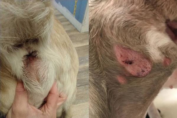
The pictures of mast cell tumors in dogs below show two of the many, many different presentations. The picture on the left shows a mast cell tumor under the skin near the dog’s anus. It looks very different from the dog’s mast cell tumor in the picture on the right, which was a raised, pink, hairless area on the side of the leg.
Mast cell tumors—the great pretenders
It is no surprise then that MCTs are know as “the great pretenders” in veterinary medicine.
I was reminded of this when my receptionist brought her Boxer, Charlie, in for a dental cleaning. She asked me to take a look at a new lump on Charlie’s side while he was there. Although the lump looked like a simple skin tag, we decided to aspirate it to be on the safe side. I’m so glad we did because it turned out this “skin tag” was actually a cutaneous mast cell tumor.
I was able to remove the tumor at the same time we did the dental. Thankfully, histopathology results indicated the mast cell tumor was low grade and completely excised so Charlie didn’t need any follow-up treatment.
I tell you this story to demonstrate how easy it can be to mistake mast cell tumors for a skin tag or lipoma in dogs. Plus, mast cell tumors may grow and shrink in size, fooling everyone into thinking they are an insect bite. For these reasons, pet owners (and sometimes veterinarians) may accidentally dismiss a mast cell tumor as being harmless. This is especially the case with well-differentiated MCTs.
Poorly differentiated vs well-differentiated mast cell tumors in dogs
When I use the term “differentiated,” what I am referring to is how much the tumor cells resemble and act like the original cells. All tumors are composed of neoplastic (i.e. cancerous) cells which originated from normal healthy cells. The more a neoplastic cell has changed from its cell of origin, the less well differentiated it is. Poorly-differentiated tumors tend to have cells that are harder to recognize, divide faster, and act more aggressively.
When it comes to mast cell tumors, well-differentiated tumors are slow growing. They may be present for months to years before they cause irritation to the surrounding tissue. As mentioned, without diagnostic testing it is easy to mistake them for a benign skin mass.
On the other hand, poorly-differentiated mast cell tumors usually grow much more rapidly than well-differentiated ones. The poorly-differentiated tumors also tend to degranulate more frequently, so the area around the tumor may be bruised or swollen. Aggressive tumors are also more likely to burst and start bleeding.
Single vs multiple mast cell tumors
Mast cell tumors are usually solitary tumors. But 11 to 14% of dogs will have more than one mast cell tumor at any given time. For example, a dog can have a well-differentiated mast cell tumor and a poorly-differentiated mast cell tumor on different areas of the body. Although they are there at the same time, these tumors are unrelated to one another.
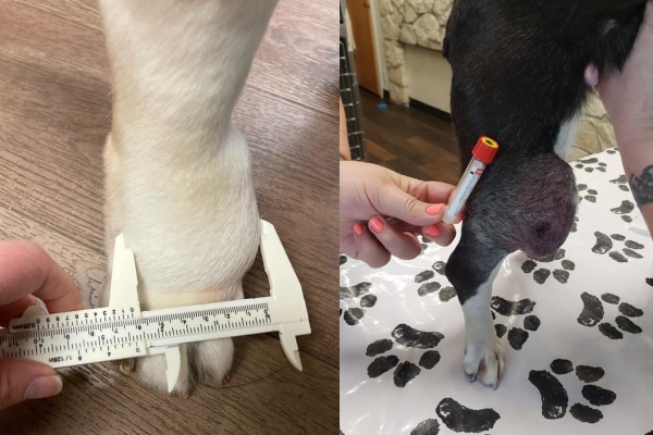
In the dog mast cell tumor pictures below, both dogs have a single, large tumor.
Spread of mast cell tumors to other areas of the body
In rare cases, mast cell tumors in dogs may disseminate, spreading rapidly and growing throughout the body. The most common locations for spread include the dog’s lymph nodes, liver, and spleen. Additionally, some dogs may develop peritoneal effusion (i.e. fluid in the abdomen, giving a pot-bellied dog appearance) as a result of MCT spread.
What are the causes of mast cell tumors in dogs?
At this point, the cause of mast cell tumors is not well understood, but there are some theories. It is possible that chronic inflammation in the skin or exposure to irritating compounds may precipitate tumor formation. Plus, mutations in a specific gene, KIT, occurs in 10-45% of mast cell tumors in dogs. This mutation may cause mast cells to multiply out of control, leading to an MCT. Additionally, several other genetic mutations or genetic abnormalities might be tied to mast cell tumor formation.
Which dogs are more likely to develop mast cell tumors?
As we have established, mast cell tumors can look like anything. They can also occur in any dog of any age. While they are more common in middle-aged to older dogs, I have diagnosed several mast cell tumors in puppies under a year of age. There is no sex predilection. This means mast cell tumors occur equally in male and female dogs and spayed/neutered or intact dogs.
However, there is thought to be a genetic predisposition to developing mast cell tumors. This is because they are much more likely to occur in brachycephalic dogs of Bulldog descent such as:
- Boxers
- English Bulldogs
- Pugs
- Boston Terriers
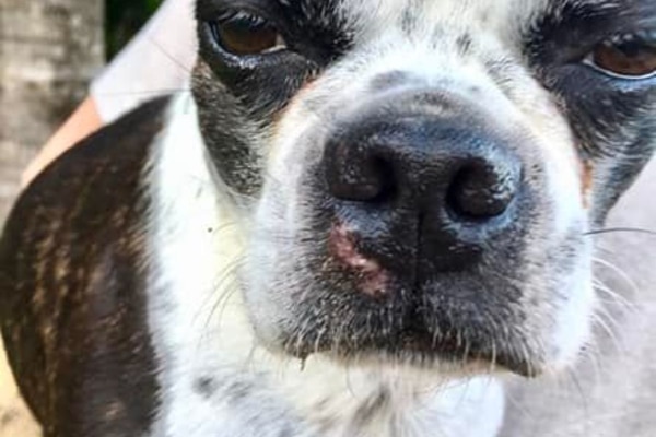
The picture below shows a mast cell tumor on the upper lip of a Boston Terrier. In this case, the dog’s mast cell tumor presented as a pink, hairless area that really didn’t look that much like a tumor at all.
Mast cell tumors are also more common in:
- Labrador Retrievers
- Golden Retrievers
- Shar-peis
- Staffordshire Bull Terriers
- Jack Russell Terriers
How is a mast cell tumor diagnosed?
Whether your dog is on the above list of predisposed breeds or not, you need to be aware of mast cell tumors. And if you only take one thing away from this article, I hope it is this—if you find a mass on your dog, don’t ignore it. One of my friends, who happens to be a well-known veterinary oncologist, has a saying, “Don’t wait—aspirate!”
Fine needle aspiration
What does the “Don’t wait—aspirate!” message mean? If you or your vet find a new mass on your dog or if an existing skin mass is changing in size, color, or consistency, the best course of action is usually to start with having your vet perform a fine needle aspiration (FNA).
During an FNA procedure, the vet will insert a 22-gauge needle into the tumor and redirect it multiple times. This fills the shaft of the needle with tumor cells. Then the vet will transfer the cells to a slide and spread them out. After letting the slide dry and staining the material, the vet will examine it under the microscope.
The video below shows a veterinarian performing a fine needle aspiration on a lump that turned out to be a lipoma in dogs and not an MCT.
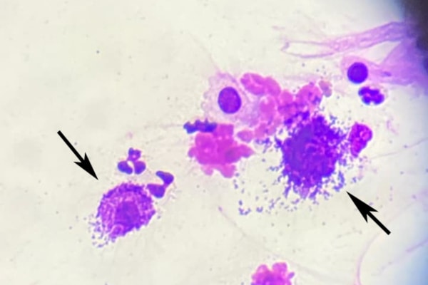
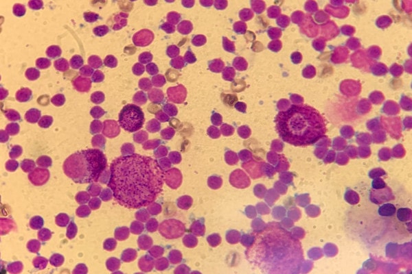
Your veterinarian may be able to diagnose a mast cell tumor in the clinic, especially if the cells have a typical appearance. Mast cells are normally round with dark-staining granules.
In some cases, the granules may not be visible with Diff-Quick (the typical stain the veterinary team would use). If your veterinarian suspects a mast cell tumor but doesn’t see the typical granules, he or she may recommend submitting the slides to a pathology lab. These labs have special stains to better identify mast cells.
Also, sometimes the veterinarian may not suspect an MCT initially. The FNA may look like an innocent skin mass, or a pocket of infection, especially if the granules don’t stain. In those cases, histopathology (i.e. biopsy) of the mass after surgical removal will reveal that the mass was actually a mast cell tumor.
Appearance of a mast cell tumor on FNA
The image below shows how mast cells in dogs may look under a microscope.
Histopathology
If the results of the fine needle aspiration show mast cells, your veterinarian will likely recommend removal of the tumor. After the tumor is surgically removed (which we will discuss later in this article), your veterinarian will probably suggest submitting the tumor to the laboratory for histopathology. Histopathology (i.e. biopsy) is the microscopic examination of all or part of the tumor tissue.
Clients often ask me why it is important to do histopathology if the FNA already indicated this was a mast cell tumor. I understand their concern because histopathology is an added expense. However, histopathology is the next step in the diagnostic process, and a very important one.
Histopathology can provide information to help determine the prognosis and next steps for your dog.
The FNA and biopsy give different information about the tumor
To help dog parents understand the distinction between cytology and histopathology, I like to use the analogy of a brick wall. Cytology is an examination of the bricks alone. Based on the FNA, the vet can often identify the “bricks” as mast cells.
At this point, the vet knows he or she is dealing with some sort of mast cell tumor. Since this is a type of cancer, the vet will need to take wide margins around the tumor to maximize the chances of removing all the tumor cells. If the “bricks” were something benign, the vet wouldn’t need to remove as much tissue around the tumor.
What the vet doesn’t know by looking at the bricks is what sort of structure they are part of. This is where histopathology comes in. It is the examination of the entire structure—the bricks, the mortar, and how they are arranged relative to one another to create a wall.
In other words, histopathology gives the information about the structure and appearance of the tumor as a whole. And it allows the pathologist to calculate the mitotic index (i.e. the number of cells that are dividing). This information can help predict biological behavior of the tumor and is necessary for determining the grade (and therefore prognosis) of the mast cell tumor.
Grading systems for mast cell tumors in dogs
Veterinary pathologists grade mast cell tumors using two different systems.
The older system, the Patnaik grading system, evaluates mast cell tumors on a scale of one to three.
- Grade I—well differentiated and surgical removal is often curative.
- Grade II—intermediately differentiated tumors. Some tumors behave somewhat aggressively while others can be cured by surgery alone.
- Grade III—poorly differentiated tumors that tend to be more aggressive.
There have been a lot of inconsistencies with this grading system, with most tumors fitting into a Grade II category. As a result, many pathologists now use the two-tier system. They will classify the tumors as either high-grade or low-grade. High grade tumors carry a worse prognosis because they have a higher rate of metastasis and a shorter survival time.
To reach a final consensus about grade, sometimes it is necessary for the veterinary pathologist to use additional stains and conduct further testing on the tissue samples. These special tests are most frequently used on Grade II tumors or tumors that have some characteristics of high grade and some of low grade.
Staging of mast cell tumors in dogs
Additionally, veterinarians or veterinary oncologists may classify mast cell tumors into different stages depending on the number of mast cells tumors and whether or not the mast cell tumor has metastasized to other locations. In order to gather the necessary information for staging, the vet may recommend an FNA of lymph nodes near the tumor and/or an abdominal ultrasound to look for signs of metastasis. Occasionally, the dog may also need a bone marrow aspirate.
One potential staging system for mast cell tumors in dogs, which is based on the World Health Organization staging guidelines, is as follows:
- Stage 0—no spread to lymph nodes and the dog had one tumor which was excised from the skin
- Stage I—no spread to regional lymph nodes and there was one tumor confined to the skin
- Stage II—tumor has spread to regional lymph nodes but there is only a single tumor confined to the skin
- Stage III—tumor is large and deeply invading surrounding tissue or dog has multiple MCTs with or without spread to the lymph nodes
- Stage IV—tumor has spread to distant locations
Each of these stages can also be broken down into a “substage a or b” depending on if the dog is showing clinical signs. Dogs in “substage a” are not showing any signs from the mast cell tumor, while “substage b” dogs are clinically ill.
What is the treatment for a mast cell tumor in dogs?
In order to use histopathology to confirm the tumor is a mast cell tumor and grade the tumor, the veterinary pathologist needs part or all of the tumor. Plus, the end goal is to get rid of the tumor completely. So it’s no surprise that the best option for getting mast cell tumors treated is often surgery.
Surgical removal for a mast cell tumor in dogs
Prior to surgery, the veterinarian may recommend you give your dog medications to help decrease the risk of the mast cell tumor degranulating and causing adverse effects. These may include Pepcid for dogs (i.e. famotidine) and Benadryl for dogs. (But please note that using famotidine and Benadryl for mast cell tumors in dogs is not going to treat the tumor itself. It just helps protect the dog against the effects of tumor cell degranulation.)
Additionally, the vet may recommend blood tests for dogs such as a blood chemistry and CBC (complete blood count) to assess the dog’s overall health and fitness for surgery or other treatments.
During surgery, the surgeon will most likely want to take wide margins around the tumor to try to ensure that he or she removes all cancerous cells in the region. One recent study in The Journal of Veterinary Medical Science investigated using margins proportional to the mast cell tumor diameter. This is in contrast to the other approach of using a standard number of centimeters for a tumor of any size. Since there currently is not a consensus on the best approach, your veterinarian will do what he or she thinks is best for your dog’s situation.
Additional surgery, radiation, or chemotherapy
In cases of low-grade tumors, surgery is often curative long-term as long as the margins are “clean.” The term “clean” means that histopathology indicates there are no microscopic traces of the tumor extending to the edges of the incision.
If the margins are “dirty,” the excision was incomplete and tumor cells remain. In those cases, the vet may recommend a second surgery.
If there isn’t enough tissue for the veterinarian to feel comfortable performing a second surgery, he or she may recommend seeing a board certified surgeon or surgical oncologist. (Need help finding a specialist or understanding what they do? Check out my article on veterinary specialists.) Alternatively, the vet may suggest radiation therapy, which is the treatment of choice to kill the remaining tumor cells.
With high-grade mast cell tumors, or if there is evidence of metastasis, your veterinarian may also recommend a post-surgical consultation with a veterinary oncologist. The oncologist can help you determine if follow-up chemotherapy or other treatment options are best for your dog and you.
Treatment for non-surgical mast cell tumors in dogs
Consulting with a veterinary oncologist is also beneficial when surgery isn’t an option. Some MCTs are too big to be able to be completely removed. Or the tumor location may be such that surgery isn’t possible. In those situations, radiation therapy, chemotherapy, or targeted therapy may be good alternative options.
Palladia®
Some mast cell tumors will respond to the medication Palladia (toceranib phosphate), which is a tyrosine kinase inhibitor. This is an oral medication which blocks signaling through the KIT receptor, a receptor which may be mutated and more active in mast cell tumors. While it is a good option for some dogs, not all mast cell tumors respond to Palladia. Also, some dogs experience GI side effects such as nausea, vomiting, loss of appetite, or diarrhea.
Stelfonta®
Recently, Stelfonta (tigilanol tiglate injection), a new and exciting treatment for mast cell tumors, has become available. Unlike other treatment options, the veterinarian will inject this medication directly into the mast cell tumor. Stelfonta works by activating a protein that causes the tumor cells to die.
This is an innovative (if not miraculous) treatment that appears to be an effective alternative to surgery for masses that meet certain criteria (size, location, and absence of metastasis, etc.). Talk with your veterinarian to determine if your dog may be a candidate for Stelfonta.
Prednisone
The steroid prednisone for dogs may be part of some chemotherapy protocols. It also has the ability to shrink mast cell tumors temporarily in some cases. However, prednisone alone is inferior to other true treatment protocols as results tend to be short-lived. Overall, using prednisone as the only treatment for a mast cell tumor in dogs has a 20% response rate and has not been shown to increase survival time. Plus, some dogs experience significant side effects from the prednisone.
What is the prognosis for mast cell tumors?
The goal of any of these treatments is to give a dog the best possible chances of survival and a good quality of life. A dog’s overall prognosis and life expectancy are based on a variety of factors.
Life expectancy of dogs with mast cell tumors
As mentioned, histological grade plays a large role in long-term outcomes (Note: grades are listed as “Patnaik system/two-tier system)
- Grade I/low-grade mast cell tumors—95% can be treated successfully with surgical excision alone
- Grade II/low-grade—Mortality rate is 17%
- Grade II/high-grade—56% mortality rate
- Grade III/high-grade—median survival time was 108 days and only 16% of dogs were still alive one year after diagnosis
In addition, the mitotic index of the tumor cells (i.e. number of cells dividing) can also help predict life expectancy. One study demonstrated that dogs with a mitotic index of less than 5 for their mast cell tumor had a median survival time of 80 months. This means that 50% of the dogs were still alive 80 months after diagnosis. On the other hand, dogs with a mast cell tumor with a mitotic index of greater than 5 only had a median survival time of three months.
Other factors influencing prognosis
In addition to histologic grade and mitotic index, location can also be prognostic. Tumors on the prepuce or inguinal area, nail bed, mouth, and perineal area are often more aggressive. Additionally, mast cell tumors that occur internally, such as in the GI tract, liver, spleen, or bone marrow, carry a very poor prognosis.
Finally, the breed of the dog can also affect the prognosis. Remember I said that dogs of Bulldog descent are more likely to develop mast cell tumors? Well, the good news is that tumors in these dogs often tend to behave more benignly and are less likely to cause serious disease than in other breeds.
When do you stop fighting mast cell tumors in dogs?
As you can see, the prognosis and life expectancy can vary significantly. For some dogs, a mast cell tumor is just a one-time problem that surgery can quickly and completely resolve. But other dogs aren’t so lucky. Maybe they have an aggressive tumor which is causing local or systemic effects. Or they have one that is difficult to remove surgically or requires additional therapies. In those situations it can be hard to know when you should stop fighting the mast cell tumor.
I can’t answer the question of when to stop fighting mast cell tumors for your dog. But I can help direct you to some people who might be able to give you support and a bit of clarity. First of all, your veterinarian or veterinary oncologist are great resources. Like you, they are committed to helping provide your dog a good quality of life. They can help you evaluate your dog and discuss options (if there are some) for improving quality of life. Or they can help you know when it is time to say goodbye to your dog and prepare for your dog’s euthanasia.
You could also consider consulting with a veterinarian who specializes in dog hospice care. These wonderful individuals can help fill your dog’s last days with dignity and comfort. And they too can help you decide when to stop fighting mast cell tumors.

Don’t Wait—Aspirate!
Mast cell tumors are very common in dogs. But they can also be very challenging because they can look like almost any other tumor. If you find a new mass on your dog or one of your dog’s existing masses is growing or changing, ask your veterinarian to take an aspirate. That way you can breathe a sigh of relief that the mass is benign. Or you can start addressing the mast cell tumor while it is potentially still small and easier to treat.
If your dog does end up having a mast cell tumor, talk to your veterinarian about which treatments would be best. Thankfully, veterinarians and veterinary oncologists have an array of options at their disposal. They can discuss the pros and cons of each option with you so that together you can figure out how to get rid of the mast cell tumor while keeping your dog as comfortable and happy as possible.
Do you have personal experience with mast cell tumors in dogs?
Please share your dog’s story or some words of encouragement below.


My 10 year old chihuahua mix recently had a MCT about the size of 1/2 a golf ball on his chest/shoulder area – the vet didn’t take an aspirate, just recommended removing and was correct – received the pathology report and it is grade 2 MCT. A few days after his surgery to remove it, another spot came up on his foot – we don’t believe it was there before and they thought it was likely drainage from the surgery and had me soaking in epsom salt bath and gave an additional antibiotic and pain extended pain pills. After a week of this, it was no better and he was due to have his staples removed, so they kept him overnight and did some laser treatment and a soft wrap over night and the next day they decided it was likely a mass, not drainage and did an aspirate. It came back as MCT.
He is now scheduled for surgery to remove it and the vet is concerned since it is on his foot that they won’t have enough skin to close it and he will have to come home with an open wound, which is very worrisome. I don’t know if it is a grade 2 low or high at this point, I will be checking with the vet. So far, even though his little foot has this pretty large tumor on it, he seems to be getting around well and has not acted sick at all. I am hoping that it comes back at worst a grade 2 low.
I have also now found a new spot mid leg of his other back foot that I will have her look at and potentially remove at the same time as I think it is another tumor, but much smaller, more the size of an eraser. I am just worried that these are popping up so fast that it is being aggressive and I’m going to lose him.
If it turns out it is aggressive, we will just have to do what we can reasonably afford to do. I am just trying to stay positive for now!
Dear Michelle,
I am so sorry your boy is facing another surgery and wish there was a good way to prevent these tumors from forming. Hoping for favorable results and praying for a positive outcome for your pup. ♥
We took our 7 year old goldendoodle to the vet because she was limping and then I pointed out that she had had a “cyst” for almost a year (I’m now kicking myself for not saying anything sooner). The cyst was about the size of a black bean and hadn’t changed in size so I wasn’t concerned. Well they did sedated x-rays to see why she was limping and then did a needle biopsy as well since she was under anesthesia anyway.. the vet called and said she has MCT. They removed it today and we’re awaiting the histology results. I just can’t help but think that the reason she’s been limping is because maybe the cancer has spread. They said the xray images showed possible fiber tears in her knee but that is it. What do you think? Would they be able to see any MCT on xray? I’m going crazy waiting for the histology results!
Hi Molly,
I understand your concern for your pup, but please don’t hold any guilt over how things have played out for your girl. I would be shocked if the mast cell tumor has anything to do with the limping and knee problem. Most mast cell tumors don’t metastasize, and the ones that do generally spread to lymph nodes, the spleen, or liver. I am not sure I have heard of one affecting a joint. A partially torn cruciate ligament (ACL in humans) is much more likely the culprit. Here is a link to another article with more information: Cruciate Ligament Rupture in Dogs: The Partial Tear
Praying for favorable results from the pathologist and wishing you all the best.
My 13 year old Boxer/Staffy cross girl Penny was diagnosed with terminal mast cell tumour and given 30 to 110 days. This was 2 years ago. I have previously commented on her journey but wanted to update people to give some hope. We tried Palladia and she was terribly sick so we ceased that quickly and she has been on pain relief and anihistamines. She is aging and the disease is probably progressing (it was in her liver 2 years ago) but our regime would not change. She is developing dementia but is she at least 13 (she is a rescue). We have been so lucky. She loves life, has good and bad days and we are very patient with her and try to make every day her best day as we don’t know how long she will be here for. Things we do to help her. She sleeps on the bed now so I can monitor if she is unwell, we have blow up mattresses around the house so she isn’t on the hard ground, I make all her meals including bone broth (I think this has kept her alive) which she will always have even if unwell. I give her medication in soft caramel as she will always take it and its a treat. If I can’t control her pain, its over. Routines are a priority. I don’t go out at night as she has sundowner and needs settling. We have people over so she is not left for too long during the day. We go for walks and car rides (her favourite) every single day unless she is not well and then its a car ride to get the wind in her face and it always calms her, I turn off all noises like phone, door bell, microwave as that can wake her. If she sleeps, the house is still. She has regular vet visits to our home as she gets distressed at the vets. If she needs settling, we do that. She is our priority. I am lucky to work from home so I can do this. I can be up 3 or 4 times per night with her and at times I sleep on a blow up mattress with her downstairs so she can get outside easier and she is closer to the floor. She is worth it and still brings joy and happiness to our home. On a good day, its fantastic and on a bad day it can still be beautiful as she needs cuddles and we just hope it will pass. It usually does after 24 hours. Is it hard ?… it can be, but its worth it. There will be a time she will not be here. I will have no regrets and she is living a good life but I know we are on borrowed time. I hope this gives someone some comfort.
Dear Ali,
Thank you for sharing your story about Penny. It is clear she is dearly loved and very well taken care of. Wishing you both continued success and praying for comfort and peace. ♥
can you please share your homemade dog food recipe?
Hi, I was told that as mct are caused by “broken” histamine switches, any fna can cause massive release of histamine and cause more mcts to appear?
Hi Anna,
These are great questions! While I have never heard of a “histamine switch” or come across it in my research, that doesn’t mean that one doesn’t exist. There is a risk of causing a histamine release when performing an FNA (or surgical removal) on a Mast cell tumor. For this reason, we routinely premedicate with Benadryl to try and prevent a histamine release from occurring. Thankfully, this histamine release does not cause more MCTs to appear. Research shows that dogs with an MCT have an 11-14% chance of developing additional MCTs regardless of whether an FNA is performed or not. If your dog has a mass that needs to be investigated and there is suspicion that it could be an MCT, I recommend moving forward with the FNA and just having your pup premedicated prior with Benadryl. Hope this helps a bit. Wishing you and yours all the best!
Our 14 yr old hound/boxer mix (77 lbs) who already had some arthritis in his back legs ad surgery to remove a mast cell tumor and was doing quite well for 5 days following surgery. In good spirits and eating and walking normally (his usual a little stiff, but not wobbly). We limited activity to just bathroom breaks and minimal walking around yard with lots of rest.
He was already on 75 mg of carprofen once a day. Post-surgery, the vet upped his carprofen dose to 75mg twice a day and added gabapentin (low dose 300 mg) twice a day. After the 3rd day we discontinued the gabapentin as he seemed to not be in any pain. He was eating well and able to walk without difficulty for 2-3 full days afterwards.
Then, in early morning hours of the 6th day post-surgery, he woke us up with very wobbly pacing. He’d wobble a few steps and then partially collapse as his back leg would give under him. This persisted over the next day and we took him to vet for follow-up. Blood work came out fine, and after a physical exam the vet said it could be from post-op inflammation as well as some lower back vertebral pain that she had detected. He was given a methadone shot and increased his gabapentin to 600 mg twice a day.
But for the past day and a half since his follow-up, his wobbliness has gotten worse to where he can barely walk. Though he still will walk to his food dish and eat normally and can walk enough to pee and poop on his own…with a lot of stumbling. His stool looks normal.
We’re wondering over the weekend (with no access to vet counsel) if such a sudden onset of wobbly gait (ataxia?) can occur 5-6 days after MCT surgery? And if so, is this severity of it common? Is it usually transient. and if so, how long might it take to pass. We just don’t know how concerned to be as he otherwise seems like his normal self.
Dear Stacey,
I am sorry your boy is experiencing these worrisome neurologic symptoms. While I can’t say for certain that this is unrelated to the MCT surgery, I cannot come up with a good reason for how they would be connected. Rarely we do see dogs throw a blood clot after surgery (of any type) that can cause stroke like symptoms, and it is possible your pup has experienced a worsening of his back pain. But one thing to know is that gabapentin can cause ataxia and wobbling (sometimes to a severe degree) especially when first started. It tends to be dose dependent so if you lower the dose then the symptoms get better. Also, if you give a dog some time for their body to adjust, the wobbling seems to resolve on its own even if you continue the medication. Here is an article with more information: Gabapentin Side Effects in Dogs? A Veterinarian’s Answers
Hoping you can find the answers you need and praying for a full recovery for your sweet boy.
I read the prior text and concerns about the dog becoming wobbly post up. I had that happen with my dog who unfortunately passed a few years ago. she went in for a simple dental and did horrible with anesthesia and that when I brought her home she was mentally off and wobbly and I did not know why. this continued and about 6 months later I had a second vet look at her and that was when they discovered she had cushions disease. it only presented after the anesthesia postoperatively. it tweaked something the vets do not know what I’m just cautioning you to make sure that you mentioned that to a vet if you take your dog who is still struggling with this because it happened to us! and that was a very hard Road until I had to have her euthanized and broke my heart. she had no symptoms prior to this Dental procedure whatsoever!
Hi,
Thanks so much for sharing your experiences with your own dog becoming wobbly after her dental. I’m so sorry to hear that you ended up having to say goodbye to her and that her last months were difficult. Your dog’s story is a good reminder that unfortunately, dogs don’t always do things “by the book.” Sometimes we don’t know, and may never know, why things happen the way they do. I appreciate you taking the time to share this word of caution with other dog parents.
My 8 year old Boston Terrier, Fitz, was diagnosed with a MCT (Stage 2) in July of 2024. It was on his outer hind leg/thigh. They were able to take out the tumor but not the margins due to the skin being so taunt there. They suggested going back in to take the margins but that would have required tie over stitches which was essentially an open wound. I declined because of the risks of infection. X-rays indicated it was in just that area but maybe in lymph nodes too. They gave him 4 months to live. It’s been 15 months and he just recently developed a 2nd bump the size of an eraser this time on his head above his eyebrow. Just got the call it was positive for MCT. I’m unsure what to do. Part of me thinks that removing it could increase his life expectancy just like last time it maybe did. The other part thinks maybe it is his time to go soon anyway and getting this surgery would just make his last bit of time not enjoyable. I adopted him in October of 2023 after he was abandoned and abused most his life. I just feel so selfish because I want more time with him. On top of everything he seems so healthy and happy as is, so it’s hard to believe he is so sick. I’ve barely had him with me for 2 years . He is so silly and kind, kissing away my tears as I write this. I’m not ready to lose him.
Dear Kay,
My heart goes out to you as you face this difficult situation with Fitz. I understand why you are conflicted and wish I had some good advice to offer. Unfortunately, there is no right or wrong way to proceed, and you have to trust your intuition as you know your boy better than anyone. It is ok to forgo surgery and choose comfort care to focus on quality of life. Either way, I am certain Fitz knows how much you love him and that he was lucky to find you when he did. Praying for clarity and strength as you navigate this emotional path. Bless you and your sweet boy.
It may not be standard treatment but there is at least one veterinarian who advertises that they simply freeze off these bumps just like they were warts on a human being. Of course they receive free medication to keep them from having a terrible side effect if the mast cell tumor releases Substances that can cause An allergic type crisis(degranulation). but the doctor claims success and Being able to do small bumps sometimes with no anesthesia but other times just enough sedation to make sure they hold still so the aim is good period since that is a licensed veterinarian it seems worth investigating if for some reason you and your regular vet don’t think you want to put the dog through the extensive impact of a surgery but you don’t want to just lose him because nothing is done. Your vet may or may not know about this other person Or have an opinion about it but it wouldn’t hurt to ask.
Should have Proofread my comment better before sending it. I said of course they receive PRE-(not free)-medication before the procedure, because disturbing a mast cell tumor causes it to release histamines and all sorts of substances that can cause problems elsewhere in the body like gastrointestinal bleeding. So they have to be premedicated with antihistamines, stomach medicine, etcetera. But the treatment with the freezing is not a fringe bonkers idea. It’s just not been heavily researched and so is not standard and many vets would not consider it for that reason and there’s not a lot of information to know statistically how well it works. However it doesn’t just one vet doing it. That’s pretty much all I know except that if I weren’t going to do any kind of surgery and that vet were within driving distance of me I personally would absolutely have it done because it couldn’t do more harm than ignoring the cancer unless it caused spread or unless the premedication was inadequate and disturbing it caused a crisis. Because there isn’t a lot of research I imagine there’s not much information about whether it can spread but the The fact that you’re freezing the tissue at the same time as you’re killing it probably means it would be hard for it to release much into the bloodstream before it froze solid because he uses the same type of liquid nitrogen with applicator that they use on boards. (Yes, they do sell DIY wart freezing kits in the drugstore for consumers to use on regular warts, but please, people– don’t even THINK about doing that on your own. You would not be able to do it in a way that would not ’cause that degranulation and you could end up causing your dog to die! Plus getting the depth right so that it destroyed the bump without going too deep would definitely be something only an experienced vet would be able to do. I’m just saying this clearly because I know the temptation would be there If you’d heard of wart-freezing kits, and in no way would that be a good idea!)
That’s just my opinion on my dog though–the choice to try that vet if surgery was not a reasonable option or unaffordable. I was lucky enough to get Stelfonta for my dog. She had a mast cell tumor that was operated twice and returned twice and even spread to the lymph nodes in the groin although only a single node. Stelfonta not only removed the tumor completely and permanently without causing any more scarring of her poor little thigh, but also destroyed the cancer in that single contaminated lymph node. She lived another 4 years without a recurrence, which was 5 1/2 years or more from the first tumor appearance. That really was miraculous considering it was a mid level aggressiveness tumor. But it can only be used on certain places like the legs and is expensive and not all vets even know how to use it. Veterinary teaching hospitals do though and it’s definitely worth calling around.
Hi BostonMominMO,
This is definitely the first I have ever heard of trying to freeze off a mast cell tumor. While my intuition tells me this is probably a bad idea, I can’t say for sure as I have not seen any research or heard of any firsthand accounts of this being done. It is an interesting topic for sure. Thanks for sharing with us! Best wishes to you and yours.
My thought is as long as it’s happy and living well (sleep, eat, good energy), it should live. Euthanasia does not exist in a dog’s world. Don’t use it lightly. Especially your dog has not been lovingly cared for before. Give him a chance to experience happy life. There are antihistamine and stomach acid suppressant and pain meds that vet can prescribe. Like human, no one will opt for assisted dying when asymptomatic and no doctor will approve.
It is January 17th 2026
I have a pit bull named Alice. I had noticed she seem to be not feeling very good, and told my boyfriend that i think she might be a Lil sick and not feeling very good or something. Maybe about a week later or so, On Sunday December 14 I went to give my dog a bath, she had been wearing a sweater jacket on the count she hates being cold and we live in Colorado, I took off her jacket and started to the water to get it warm enough for comfort, as my dog was sitting there I noticed a big mass at the base of her neck more to the left side where her breast plate is beginning next to her throat. I thought good lord where in the heck did that come from out of no where, how come I didnt notice this sooner, as this seemed huge to me and was sticking out. One would have to be blind to not see it. but it popped up out of nowhere. I told my boyfriend to come look. The soonest I could take her to the vets was Tuesday December 15th 2024. So we wnt in and saw the doctor, she told me it was cancer. and we talked about setting up an appointment to do an x-ray and blood work cuz she is 9 years old to make sure she was well enough for surgery to remove it. How every they were all going to be out of the office till after the 1st of the year for the holiday’s. She gave me a caliper to use to keep track on how fast it was growing. and told me to schedule her an appointment for what we had talked about. She decided that aspirating it with a needle that she wouldn’t be able to draw any thing back out of it. She mentioned that she thought it could be a hard surgery do to the location, as it tucked down next to the beginning of he breast plate and wasn’t sure how much it could be going down inside her chest, or if she would be able to get it all out for sure. So I never been through anything like this and it was all new to me, and it all caught me off guard. I went home and spoke to other family members about it a bit. I wanted to get her in ASAP for the x-ray wanting to make sure weather or not it has spread, before I decide to put her through all of this. But would now have to wait till they were all back from the holiday as this vet is supposed to be a more affordable vet and come to my town every Tuesday, and work in another town on Mondays and Wednesdays. and couldn’t been seen in the other town on Wednesday as they were booked. So now that this had time to sink in my head for a minute, I had a lot of questions and no one to answer them. But the way she made me feel at the appointment made me feel like she had time or that It wasnt a rush right this second thing. So she had measured the mass in her office, it was like about a 4 on the caliper she gave me to use to kind of keep track on how fast it was growing. so every night I was checking it every night. it hadn’t even been a week yet and it went from a 4 to a 6 and then an 8 and I was like good lord this thing is growing to fast she’s never going to make it till they get back at this rate, and i had some concern about this thing possibly strangling her. So I contacted another vet and all he wanted to do was put her down, acted like if I brought her in and he thought I should he acted like he wasn’t going to let me leave with her, so he was definitely out for us. I happened to find a hospice vet that worked in and out of our town I had no idea they had them for pets. She did home visits, I thought great as she told me she would stop by tomorrow after noon and come take a look at her. She spent more time with me and my dog then even a human doctor spent with me. She aspirated the mass twice but was only able to get blood out. and put it on slide anyways but let me know it could come back inconclusive. said she would get back with me about the results. Gave me a couple prescription . was kind of hesitant on the one but decided to give me for her anyways, Carprofen and Gabapentin.this was on December 23. The next day I was doing something outside and my boy rush outside to find me and says to stop what I’m doing to get inside right away and Im like whats going on and he says it ruptured so rush inside, he was panicked and thought she had 3 to 5 minutes before she’d bleed out, I check her over and I thought it had maybe bled a Lil possibly from the poke in it for the day before, but hadn’t ruptured but she was ok. so I had her just lay down and sang to her so she would calm down cuz she was tripping now too. she settled down and went to sleep and was ok. The next day I had company as it was now Christmas. I stayed in bed with her beside me. she got excited and wanted to bark and run for the door every time someone would arrive but I wouldnt let her get up and run for the door. with the other dogs. my son came in the room to talk with me and see how things were going she was excited to se him. I turned my head to look at him and she decided to try to scratch he mass, she was able to get one stroke acrossed it with her foot as I grabbed and so no you don’t and stopped her. she went to the edge of the bed to see my son and he was like is this thing supposed to be leaking like this and there was what looked like a spray of blood acrossed the foot of my bed and I was like shit, I got some paper towels and started to apply some pressure and had been for a while, ever so often checking to see how much it was bleeding she had a good Lil gash in it, I I kept adding paper towels as needed. My boyfriend had been doing the dinner thing for us all and brought me a plate I still hadn’t yet mentioned what had just happened. I mean it was bleeding but she was like bleeding out or anything. I finally asked him to get me he coat and a towel so I could use her coat to hold the towel in place so I could eat while explaining what had happened. we couldn’t get the bleeding to stop completely and so my son’s girlfriend went to there house to get some bleed stop to try it as I had already messaged to hospice vet, but still hadn’t heard anything back yet. we finally got the bleeding to stop with the bleed stop and by using layers of toilet paper.. By this time company was long gone and I was finally able to try to rest a Lil bit, about 5 in the morning the vet finally messaged me back, wanted me to send her a picture after explaining everything and told her I was worried about trying to take everything off to do so as we had a hard time getting it to stop bleeding as it was and she was wanting me to at least wash it later so it hopefully didnt get infected. oi said ok even know I had no intentions of taking the tp paper off causing it to reopen. the next day she scratched again and pealedit off herself so I sent the vet her picture. it leaked a Lil bit but not to much..And the bleed stop worked well to seal it up the night before. The vet wondered about the blood loss I told her her gums were still pink and I thought she was ok. She asked if I mind her stopping by on her way through the next day I said it was fine for her to. However she had a emergency and never made it back by. After everything I’d been through I had made up my mind I did not want to take my dog in to a vets office to be put down because I was so close to this dog the last thing I was going to do was take her to a place the she smells nothing but fear and death in there and shakes like crazy to just go in to see the vet, that she is not comfortable at all at muchless does she wanna lay down a go to sleep or be force to sleep. so I decided that was not for us. I had made my mind up that I’d prefer to keep her here at home with me where she is happy and comfortable, I just wanted to be able to keep her as comfortable as I could till she passed and have her creamented and have my kids put her Ashes with me when I passed. I never expected her to make it till January 7th the day of her appointment. It had made me start thinking its great if you find this early but by the time you start showing signs of illness or sickness its already to late. The day before her appointment the vet on holiday break has someone call me, and asked me about doing the bloodwork and x-ray but going a head and doing the removal at the same time. I was like absolutely not as a lot had gone on sense they had all been on holiday and explained to her I want to know the results of the x-ray and blood work before I would agree to the rest of everything else cuz per say it had spread and she had 2 weeks left to live or something I couldnt see making her go through all this making her last days horrible. So I took her in to the appointment, mind you this dog and I had been inseparable sense finding out on Dec 15th being told she had cancer. The doctor wasn’t in yet as it was 7:30 Am when we arrived and I had never been to this other vet clinic office before in this other town. It was already packed with a full parking lot, I went in to talk to them first and was trying to explain everything that had gone on the past few weeks. But keep being interrupted with all these people and their pets. all these dogs were in kennels lined the wall in this small office right inside the door as come in. as the dogs we barking and it was pretty hectic. All a sudden the man working there tells on of the ladies to get that yappy Lil dog out of here in not putting up with this all day, take it next door to be put in another lil building equivalent to a Lil barn shed type building. and she did, only to come back and ask me to bring my dog in to get her weight.. I said ok but still wasnt able to fill them in on what was going on with my dog. then tells me to bring her to the other building where she had just took the yappy dog. I was cold in there and they kept coming in and out the door which made it that much colder. She was already a shaking mess by now and the last thing I wanted to do was leave her in there. I went back to the first building to try to talk to them about whats been going on and I ended up stepping outside to talk to this same not nice man, And he couldnt shut up long enough for me to finish explaining things to him, as I was trying to I told him about the hospice vet and the meds and went to mention that, that they did the needle poke on her mass and he starts yell at me we did not we did, said I didnt say you guys did, and he had this additude with me cause I had said I wanted to talk to the doctor before I left, and he was like maybe this isn’t the vet for you and I was so frustrated at this point that I was like ya maybe your right let get my dog then and so we did and I left. so here I sit not know what the heck I should do. he told me that they would have to do the blood work and then put my dog to sleep to do they would do her x-ray. I know another vet I could take her to to get the x-ray and they wouldnt put her to sleep to do it. and at this point all I wanted to know was has it spread already or not, before I made any other decisions, and hve been wondering if I should just do that for now. for the past so many days. which is what I will probably do this week., to start with for now. I also left a message with the hospice vet today to let her know what had happened also. I have yet to hear back from her. This has taken its toll on me this dog is my best friend and has been my emotional support after I lost my father, and this has all been so hard on me. The mass on my dogs neck has sense shrunk down quite a bit but is still there so I came to this site to find out more information about the shrinking of her mass and trying to figure out what out best options are and came acrossed this new treatment shit they can give your dog that I guess helps kill the cancer cells or something like that, which didnt sound to bad but didnt really have enough information about it. this is my story so far just looking for more information about this new shot now and and maybe a Lil advice about all this.
Dear Patricia,
I am sorry you have been through so much with Alice over the past month. While I would love to be able to offer insight and advice, it is hard to know what is best without being able to examine your pup for myself. I am not sure what new treatment you are referring to but would be happy to provide additional information if I knew the name of the medication. From what you have described, I am worried that part of the confusion you are experiencing is due to having so many different medical professionals involved and none of them being able to communicate with each other. Each vet will have their own opinion, and it is ok to disagree. Ultimately, you have to trust your intuition and do what you feel is best. You know your girl better than anyone. My best advice is to find a vet with whom you can put your trust and confidence and partner together to make a plan for how to proceed. Praying for a positive outcome for Alice and wishing you both nothing but the best.
we have a pug nearly 2 years old she is tiny and such a cute baby. but she has developed a growth looks like a white cyst in her left nostril and she has been vomiting acid up every day mostly at night time. I have a vet appointment for her on monday but I am worried
thanks
Penny
Hi Penny,
I understand your concern for your pup and agree that she definitely needs to be examined by your vet. Hoping you can get the answers needed to resolve these worrisome issues. Best wishes and bless you both.
Hello,
My dog was just at the vet today, so she hasn’t been officially diagnosed with MCT. But the vet and I highly suspect that is what we’re dealing with. I wanted to share some information that helped my dog. Granted, my experience may be anecdotal, but this did seem to help. My 9 year old English Coonhound developed a tumor on her neck, just above her shoulder. Because of where it is, cones and inflatable donuts are useless. Despite my best efforts, she was able to scratch it open and it began draining before her vet appointment. I’ve kept it bandaged and was giving her benadryl, but she continued to try scratching at it. Sunday it swelled up worse, at least doubled in size, and began draining a good bit of fluid mixed with blood. At my wits end, I tried a poultice that is typically used for horses when they have an abscessed hoof. One part Epsom salt, one part sugar mixed with enough betadine to make a paste. I applied the poultice and bandaged her neck as I had been doing and continued the benadryl. I left the bandage on for 24 hrs. She finally left the bandage alone. Monday morning during her bandage change I noticed the drainage and swelling were significantly decreased. So i repeated the process. This morning the swelling was almost completely gone and the drainage had stopped. Obviously this is in no way a cure. But it did seem to give my dog a good bit of relief. Dr. Buzby, could you weigh in and let me know if it was coincidence that this helped, or if people could use this process to help their dogs with open tumors.
Hi Cindy,
I am sorry your girl is dealing with this worrisome mass on her neck. I have definitely used betadine and sugar bandages to help with slow healing wounds on dogs. I can’t say that I have ever included Epsom salts. I have used Epsom salts to soak paws but never in a wound. Also, the wounds I use sugar bandages with are usually due to some sort of trauma and not a cancerous tumor that has ruptured. While it should be fine to use these products in this manner, I don’t have any personal experience to share. I would still advise readers to discuss wound care options with their vet before trying something new. Hoping you can get a definitive diagnosis for your girl and praying for a positive outcome.
there is a spray for horses that is Manuka honey and colloidal silver, you might try that.
also heard good things about black salve! there are FB groups on how to apply it..