If your older dog is “lumpy and bumpy,” lipomas are a common culprit. But how do you know if a new lump is just a benign lipoma in dogs or a more dangerous mass? Help is here. Dr. Jennifer Shepherd (friend and colleague of Dr. Julie Buzby) shares the causes, diagnosis, and treatment for lipomas (fatty tumors) in dogs. Also, through images, pictures and video, you’ll know exactly what to expect if your veterinarian suggests a fine needle aspirate for your dog’s suspected lipoma.

A lump the size of a softball—Is it a dog lipoma?
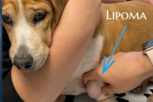
Before we get into the causes, diagnosis, and treatment of lipoma in dogs, let’s meet a 12-year-old Pit Bull mix named Maggie. Until now, she had always been in good health.
However, over the last couple of weeks, this sweet senior dog seemed relentlessly plagued by a nagging cough. Maggie’s mom was worried not only about the cough, but also about the softball-sized lump on her beloved dog’s chest that she had discovered six months prior. It appeared to be growing.
Day after day, one thought cycled through the dog mom’s mind: Could the lump on her dog’s chest be pressing into the lungs and causing the cough? Even more fundamental—what was this mass?
My conscientious client scheduled a veterinary exam with me. As I examined my patient, it wasn’t hard to find the mass. It was almost five inches in diameter, soft, and located just under the skin on top of the muscle. Although I thought I knew what the mass was and was fairly confident it didn’t enter the chest, I ordered X-rays because I knew we needed to get to the bottom of this sweet dog’s cough and congestion.
Getting to the bottom of the mass on the dog’s chest
The X-rays confirmed my suspicions. The source of my patient’s cough was bronchitis. And while the mass was there, it was on the outside of the chest wall, growing away from the chest cavity. My client was visibly relieved.
Next, to put to rest my client’s fears, it was time to investigate my patient’s softball-sized growth.
In Maggie’s case, based on the shape and feel of the mass, I strongly suspected it was a lipoma (fatty tumor). But a physical exam alone simply is not enough to determine the identity of the mass.
I knew she needed a fine needle aspiration (i.e., extraction of cells using a small needle and examination under the microscope) of the mass to rule out a mast cell tumor, soft tissue sarcoma, or other concerning tumor types and to confirm a diagnosis of a lipoma.
What is a lipoma in dogs?
A lipoma is a common benign (non-cancerous) tumor of adipocytes, which are fat cells. Lipomas usually have well-defined boundaries and typically do not invade the underlying tissue. They are commonly located on dogs just below the skin of the trunk and limbs of the dog’s body. However, in some cases, a dog may have a lipoma in the chest or abdominal cavity, or one may grow between the muscles of the leg.
Occasionally, a dog may have an infiltrative lipoma—one that invades muscle, bone, nerves, or other tissues. While benign, infiltrative lipomas do disrupt the adjacent tissues more than other lipoma types. They are especially problematic if they cause spinal cord compression or invade muscle tissue.
What causes lipomas in dogs?
It is uncertain what causes lipomas. As with many conditions, the causes are often multifactorial, with environmental and genetic factors likely playing a role as risk factors. These fatty tumors are more common in senior dogs but can occur at any age. While any dog breed may have a lipoma, the following breeds are more likely to develop lipomas:
- Labrador Retrievers
- Beagles
- Doberman Pinschers
- Miniature Schnauzers
- Cocker Spaniels
- Weimaraners
- Dachshunds
How do you diagnose a lipoma?
A physical exam is not sufficient to diagnose a lipoma. This is a critical point that I can’t emphasize enough. Your dog needs a fine needle aspiration or biopsy of the lump to be able to definitively diagnose it as a lipoma.
Even a board-certified veterinary oncologist cannot diagnose a lipoma simply by looking and feeling. A fine needle aspiration is critical to rule out other below-the-skin tumors that can be of far greater concern, such as mast cell tumors in dogs and soft tissue sarcomas.
Fine needle aspiration
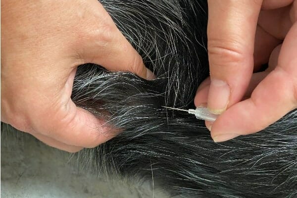
A fine needle aspiration (FNA) is a simple diagnostic test your vet can typically perform in the exam room on an alert and conscious dog. Although watching the FNA may be unsettling for owners since it involves a needle, most dogs remain calm and react to the procedure no more than they do when receiving a vaccine.
To perform a fine needle aspiration, your veterinarian:
- Inserts a needle into the mass
- Redirects it multiple times to gather cells
- Removes it with a sample of the tumor’s tissue
Watch a fine needle aspiration on a dog’s lipoma in this video…
As you can see in the next video, the vet will use an air-filled syringe to spray the collected material onto a slide, spread it out, and then examine it under the microscope.
Microscopic characteristics of lipoma in dogs
Because lipomas in dogs are mostly fat cells, the material on the slide often looks like oil. The image below shows how the lipoma aspirate looks when smeared on a slide.
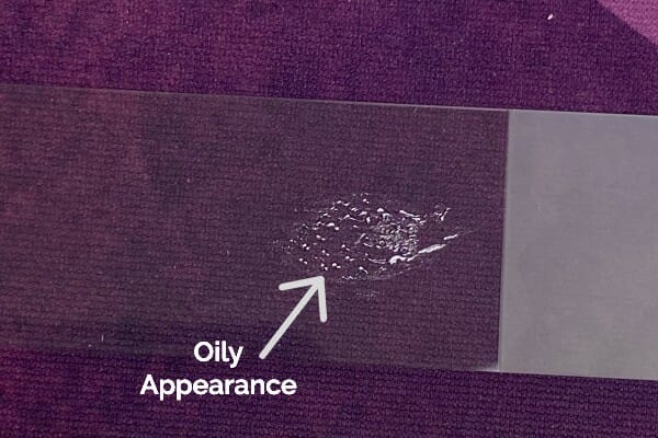
Although it may be tempting to assume it is a lipoma based on the visual appearance of the material on the slide, a microscopic evaluation is still necessary. Other tumor types can occasionally have a “fatty appearance”, too.
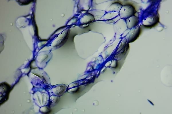
When the vet stains and observes the material under the microscope, he or she will see adipocytes (fat cells) and sometimes red blood cells if the tumor is a benign lipoma. However, if he or she sees other cell types in addition to the adipocytes and red blood cells, this suggests the tumor is not in fact a lipoma and bears further investigation.
What is the treatment for lipomas in dogs?
Many pet owners find masses on their dogs and become concerned that their dog has cancer or that the mass is going to compress vital organs. But lipomas are often asymptomatic and usually do not cause any problems with a pet’s health. The exception to this would be infiltrative lipomas and those in the chest, abdomen, or between muscles.
Lipoma removal in dogs
In some cases, lipomas in dogs can grow large and, based on location, interfere with a dog’s mobility. For your dog’s comfort, it’s best to remove these lipomas surgically. It’s important to note that the surgical removal of one lipoma does not prevent another lipoma from developing in a different area.
If the lipoma is relatively large, your vet may need to temporarily place a drain in the surgical site. This can help prevent a seroma, which is a large pocket of fluid that fills the space where the mass used to be.
It is important to follow your vet’s instructions about incision care, exercise restriction, and use of an E-collar after lipoma removal. And if you have any concerns about how the incision looks or how your dog is acting, don’t hesitate to contact your vet.
Infiltrative lipomas in dogs—aggressive treatment approach
Infiltrative lipomas often require a more aggressive surgical approach and occasionally multiple surgeries or even a dog leg amputation. If the veterinarian is not able to remove the whole infiltrative lipoma, it is likely to recur. Radiation therapy (either on its own or combined with surgery) can also be effective in controlling these types of lipomas.
FAQs about lipomas in dogs
There are common questions and concerns that many pet parents have about lipomas. You may have some of the same questions. So, let’s get answers to some frequently asked questions about lipomas.
Q: Can you shrink a lipoma in dogs?
Unfortunately, at this point, surgery is really the only way to get rid of a lipoma. There are no commonly available or practical solutions to shrink a lipoma.
One small study of 15 dogs did indicate that injecting a steroid (triamcinolone acetonide) into the lipoma under ultrasound guidance could either shrink the tumor or cause it to regress completely. Unfortunately, some of these lipomas later recurred. A paper detailing the study entitled “Canine Lipomas Treated with Steroid Injections: Clinical Findings” is available if you want to read more about it. At this point, it is not a common procedure.
Previously, some vets tried injecting calcium chloride into subcutaneous (under the skin) lipomas. However, this method has fallen out of favor because it has the potential for complications such as necrosis (death) of the skin and irritation.
Q: Are lipomas in dogs dangerous? Harmful?
A: All lipomas are benign, and most are harmless—they don’t cause any symptoms. However, if they grow in the abdominal or chest cavity or the spinal canal, they can compress other organs and cause clinical abnormalities such as neurological issues in dogs.
Occasionally, dogs may develop an intermuscular lipoma—a lipoma that grows between muscle layers. This type of lipoma most commonly occurs in the back of the thigh and causes lameness or impedes movement.
Q: Do lipomas on dogs go away?
Generally, once a lipoma is present, it will typically either stay a similar size or, more often, continue to slowly grow larger over time. They don’t usually go away on their own.

Q: Can dog lipomas turn malignant?
Lipomas are common benign fatty tumors. They are not malignant and cannot become malignant. Since they are benign, they also can’t metastasize (spread) to other parts of the body. It is important to note that this doesn’t mean that the dog won’t get other lipomas. It simply means that any future lipomas did not come from the lipomas that were already present.
Lipomas should not be confused with liposarcomas, which are very rare, malignant tumors of fat cells. Unlike a lipoma, a liposarcoma is locally invasive and often spreads to other parts of the body. You do not have to worry about your dog’s lipoma turning into a liposarcoma, as they are completely different tumors.
Q: What should I do if I find a lump on my dog?
If you find a new lump on your dog or if you notice that an existing lump is growing rapidly, you should schedule an appointment with your veterinarian. He or she will most likely perform a fine needle aspiration (FNA) to determine if the mass is a lipoma or another tumor type. Once the tumor has been classified, your vet will explain whether or not your dog needs surgery to remove it.
If your dog has multiple masses, it’s helpful to have a “body map” showing the location and size of all the masses on your dog. This will allow you to determine if a mass is new or if an older mass has grown in size. Your veterinarian will likely keep a body map in your dog’s medical record. But you can also keep one in your dog’s health journal to note changes at home.
Q: My dog is covered in lipomas, and I just found another one. Does it need to be aspirated?
In short, yes. Let me explain my answer through the story of one of my canine patients.
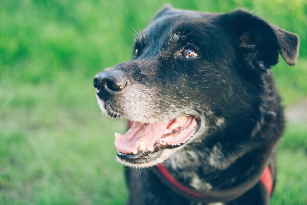
I had previously diagnosed Chloe, a 9-year-old black Labrador Retriever, with a handful of lipomas. When she came to the animal hospital for her annual exam, I found a quarter-sized mass in front of her shoulder. (This is one of the many reasons I cannot overstate the importance of taking your dog to the vet for wellness exams.)

Scanning her records from last year, I didn’t see a mass noted in this location. Although it felt like a lipoma, I wanted to be thorough.
I performed a fine needle aspiration to collect a sample. The image below demonstrates this procedure.
After I smeared the sample on a slide, I saw the classic oily appearance with what appeared to be red blood cells mixed in. Upon examination under the microscope, however, the material that looked like blood was actually a mixture of red blood cells and neoplastic (cancerous) round cells.
My patient’s new mass was not a lipoma—it was a soft tissue sarcoma, which is a malignant (cancerous) tumor. I advised surgical removal at the next possible opportunity.
This story serves as a good reminder of the importance of having your vet aspirate every new lump. Even if the first dozen growths have been lipomas, you never know when the next lump will be something concerning, where early detection can make a huge difference in the outcome.
Since this is so important, I’ll say it one more time. If you find a new lump on your dog that your veterinarian hasn’t previously documented and tested, please make an appointment with your vet promptly.
Lipomas are common in senior dogs
Lipomas are very common fatty tumors in senior dogs. Understandably, I have had numerous dog owners tell me that they are scared their “lumpy bumpy” older dogs are filled with cancer. Thankfully, more often than not, these masses are nothing more than harmless, benign lipomas and unlikely to cause any adverse health symptoms.
As always, though, make an appointment with your veterinarian any time you observe something new or “off” with your dog. Just like Maggie’s mom, it will help you put your worries to rest. Or perhaps, like Chloe’s story, it will allow for early diagnosis and treatment of genuine concerns. Either way, that vet visit and fine needle aspirate are critical.

P.S. Why lumps and bumps need to be checked by a vet
I can’t stress enough the importance of seeing your vet if you think your dog has a lipoma. In taking the videos for this blog post, Dr. Jennifer Shepherd did an actual fine needle aspiration (FNA) on one of her patients with what was assumed to be a lipoma so that we could share this procedure with our readers. Later, I received this message from Dr. Shepherd: “I may have saved a life thanks to you! I saw a dog today, and the owner said that the lumps had been looked at before. So, I assumed one was previously diagnosed as a lipoma. I decided to aspirate, just to make a video for you. Turns out it’s probably soft tissue sarcoma. I’m removing it on Wednesday.” Dr. Shepherd’s message reinforces the importance of getting your dog’s lumps and bumps checked. ~ Dr. Julie Buzby, integrative veterinarian
Have you ever found a lump in your senior dog? Was it a lipoma?
Share your experience in the comments below. Finding a lump can be scary—so let’s encourage and support one another.

My 10 year old Chihuahua has a hard lump on top of his head. The vet said it is a bone growth. It does not seem to be causing him discomfort and I have been to the Vet’s twice.
Hi J’annine,
I am glad the vet does not seem to be concerned about the lump on your dog’s head. Hoping your boy remains happy and healthy for many years to come. Best wishes to you both!
My cousin lives on a farm and has 2 golden retrievers or Labradors.
He noticed the bump on on my dogs chest area
and asked me about it.I said what it was, and he said he drained it!? I can’t do that at all.But is that what you end up doing when you remove them?
Hi Layla,
No, the process of removing a mass is much more complicated than that. If the lump is not a mass, but a fluid filled cyst or cavity, then it may be drained (although these things sometimes refill and then have to be surgically removed anyway). But with a mass/tumor, we make an incision in the skin around the tumor and then carefully dissect the different tissues apart. We detach the mass from the underlying muscle or adjacent tissues and will ligate any larger blood vessels that could be a problem. Then when closing the incision, we suture together the correct tissue layers usually requiring a 2- or 3-layer closure. This of course is a very simplistic description, and we put a lot of thought into our incision placement, work hard to prevent unnecessary bleeding, did we get the proper margins if we are concerned this is a malignant tumor, and do we have enough skin to completely close the defect once the mass is removed. Hoping this helps explain the process a bit and offers a better understanding of a mass removal surgery. Thanks for asking!
Our 4. 1/2 yo Golden Retriever/Irish Setter mix was just diagnosed with a likely infiltrative lipoma. Surgery is scheduled for August 21. He seems to be on the younger side of dogs who have been diagnosed with it. Based on his age, is he likely to get more?
Hi Wendy,
I am sorry your young dog is facing this less common type of tumor. I understand your concern and think it is great you are trying to gather as much information as possible. Unfortunately, I am not sure if the age of onset plays a factor in recurrence with this type of tumor. The studies just haven’t been done, or we haven’t come across this specific bit of data while doing our research for the article. I do believe genetics can make a dog more susceptible to certain types of diseases and conditions. It may be your boy is predisposed to forming infiltrative lipomas. Hoping the surgery is successful and recovery is smooth. Feel free to keep us updated and wishing you both all the best.
My boy (amstaff x bull terrier) had a tiny lump on his side that grew and I then noticed one on his chest. The vet checked by drawing some liquid and confirmed that it wasn’t cancerous. However since then, there have been another 3 pop up and if i’m honest I really don’t like how quick they appear to grow. I will need to go and get the new ones checked.. Thank you for sharing your story.
Hi Bianca,
I am sorry your boy is dealing with all these new lumps that have appeared. I do think it would be wise to have them examined and checked. Hoping for clear answers and an easy solution. Best wishes to you and your sweet boy.
This was very informative & helpful.
My senior dog, she is around 18 & over the last year this spot keeps growing. She has no pain. Still walks around, she actually lays on it.
No vet will operate because of her age. I just don’t know what else they may be able to do for her.
Hi Kelcey,
I understand your concern for your senior girl with this large mass she is living with. Sometimes due to the size or location of a mass, it can make removal impossible or very risky. While age alone it not necessarily a reason to avoid anesthesia, age can cause other biological changes to take place that would cause a dog to be a poor anesthesia candidate. This is why I always recommend pre-anesthetic lab work to see how the internal organs are functioning prior to surgery. Without playing a personal role in your pup’s medical care, it is hard to offer suggestions or specific options. I hope you can find the best way to maintain her quality of life. Wishing you both many happy days ahead.
Hi Doctor
Our 16 year old poodle/shtz mix a what we believe is Lipoma soft ball size down penis area. Or doctor says that he is too old for surgery but it’s interfering with him going up steps..
Some are suggesting putting him down..but we just can’t..
Hi Jose,
I understand your concern for your pup and am sorry he is living with this large tumor. Even if making a choice about euthanasia is not urgent, it can still be a loving option if your boy is suffering and struggling daily. Here is a link to another article with more information on how to evaluate quality of life: Using a Quality of Life Scale for Dogs
Praying for clarity and strength as you navigate this emotional path. Bless you and your sweet boy.
my older female dog I just found one on her chest I hope it is not anything life threatening because this area i live in offers no affordable animal care.
Hi Roxanne,
I am sorry you are having a difficult time finding affordable veterinary care in your area. Praying that this new lump on your senior girl is nothing to worry about and hoping you can get her the medical attention she needs. ♥
This was very informative. Our small dog has a large lipoma sitting right next to her vulva. It began small as you indicated and has continued to grow; currently about the size of a golf ball.. Being that she is roughly 9 lbs, you can easily see it protruding as she walks. But it has not seemed to hinder her mobility. Our vet has advised against surgery, indicating that due to its location (so close to the vulva) it would not be safe for the incision and would likely get infected. However, I am of course nervous as it grows. I’m wondering is there a cap off – meaning does it reach a point and stop growing or will it just continue to grow always? I know if it does continues it will get to a point where mobility will be an issue for her back leg but right now she is okay… Thus what brought me to find this article as I continue to seek advice/guidance.
Hi Pash,
I am sorry your little pup is dealing with this worrisome mass near her vulva. While the location isn’t ideal for surgery, it still may be the best option. There is no limit to how big the mass will grow. You might want to talk to your vet about a referral to a specialist to get more information and see what treatment/surgery options are available. Hoping you can find the answers you need to make the best choice for you and your girl. Praying for clarity and many happy days ahead.
My senior rescue Portuguese Podengo had her first rabies vacine six weeks ago and her health has gone down hill rapidly. She has large lumps in her throat and shoulder and between her armpits. They are groing rapidly. She seems to get new ones almost every day and within days grow to the size of golf balls. We do not know her age but the vet guessed at about 12 or 14 years old. Her weight has dropped off her and she is feeling very poorly. She is going to the vets later on this morning and I fear we may not be bringing her home again with us. Is reactions to rabies injections at all common?
Dear Maggie,
I am sorry your senior girl is facing this scary situation. Without examining her myself, it is hard to make specific conclusions. From what you describe, I am suspicious the lumps could be enlarged lymph nodes. If this is the case, then cancer (lymphoma) would be a major concern. This is not a typical reaction to a rabies vaccine but because vaccines stimulate the immune system, it is possible to see strange symptoms in rare instances. I hope your vet was able to offer some answers and there is a clear plan for how to proceed. Praying for healing and a positive outcome for your sweet girl.
my 5 year old lab has a limpoma on top of his next he’s scheduled for removal in 3 days Friday he was running under the trees cut a small cut top of limpoma very minimal bleeding but later that night a small piece of tissue formed over the cut instead of a flat scab its a bubble type scab no drainage noted I clean it and apply basatration
Hi Dianne,
I am sorry your Lab has caused an injury at the site of the mass. From what you describe, it sounds like his body may be producing granulation tissue to fill in the defect. Hopefully this will not prevent the surgery from taking place as planned. Praying for a smooth procedure and an easy recovery for your boy. Best of luck to you both!
I have a 14 year old standard poodle. Yesterday in the after I discover that he had two lumps on either side of his neck. I live 175 miles from his vet. I decided to massage the lumps (which were not solid). I opened his mouth and massaged both sides of his gums.
He looked like he had the mumps and would not stop licking himself. I gave him water which he drank and kept massaging thru the night.
This morning the lumps had gone down substantially with one almost gone and he was no longer licking himself.
He ate breakfast and drank more water then went into the back yard where he did both numbers.
It is now midday and the lumps are almost gone and he no longer is licking himself.
Yay – problem solved!
Sometimes just common sense and patience will do the trick!
He has been a very healthy dog and seems to be fine.
Hi Rita,
I am glad the lumps have decreased in size and your pup seems to be feeling well. Of course, without examining your pup myself, it is hard to make specific conclusions. But I am concerned that the lumps you noticed may have been swollen lymph nodes. If this is the case, it would still be a good idea to have your boy evaluated by your vet. Reactive lymph nodes can rapidly change in size and just because they shrink doesn’t necessarily mean all is well. Wishing you and your sweet boy all the best. Keep up the good work!
What is the approx cost for this type of exam?
Jill
My terrier mix is 13 years old. I rescued her at 2 years old. After a few months, I noticed a small lipoma on her chest. It was aspirated, and growth was measured often. It is a lipoma. Now it is the size of a melon. It was left to grow larger at every measurement. Now she is too old for surgery. Should this have been dealt with when the lipoma was small, when surgery certainly was not as risky.?
Hi June,
I am sorry your senior girl is living with this large mass on her chest. It is always easier to remove lumps while they are small, but it is hard to predict how they will change over time. I understand your concern with anesthesia, but if removing the lipoma would offer her a better quality of life, I would not use age alone as a reason to forgo surgery. Here is a link to another article with more information: Is My Dog Too Old For Anesthesia?
Hoping you can get the answers you need to make the best choice for your pup.
Hi Dr Buzby, I have a 13.5 year old American Pit Bull Terrier who has a lump in the back right side, lower area of her neck (pretty close to her shoulder). It is for the most part soft and does not hurt her to touch. It is completely under her skin. It is about the size of a half tangerine and has been that way for a long time. While I know it’s ridiculous to ignore the situation because of fear of the diagnosis, is it necessary to bring her to get that checked? It does not hurt her, she is in exceptional health for her age, and has no real concerns that aren’t normal for a dog her age. I likely will not choose to do an invasive and risky surgery on her because I want her to live her life in happiness and pain free and don’t want to ruin the precious time she has left with me (I did this in the past and while I don’t regret trying to help my cat, she had no idea she was even sick until I messed with her and she declined RAPIDLY – I can’t do that to my dog 🙁 it is just not fair to her..) Do you think that her lump could be lipoma if it is not outside her skin? I will ask her vet to aspirate at her next appointment in the fall. Is there something I could do (maybe fish oil?) to try to help the size go down at all? I love her with my whole heart and will do anything to make sure she has a happy rest of her golden years, so any thoughts would be welcome. She is my best friend and I am not prepared to say goodbye to her. Thank you!
Hi Alix,
I understand why you are worried about your girl with this unknown lump on her neck. I wish I could offer some reassurance, but without doing some testing, there really is no way to know what this mass could be or if it is cause for concern. Fish oil is a great supplement and does wonders for skin and joints, but I am not aware of any beneficial effects it would have on a mass. I think your best course of action is to schedule an appointment with your vet. Even if you choose to forgo treatment, it may bring peace of mind to know exactly what you are facing. Hoping for answers and a clear path forward. Wishing you both all the best for many happy days ahead.
thank you!
I have a 5 year old white Westie/ Maltese guy named Augustus. (aka”Gus”)
He has a lipoma on his tail. High up towards butt. Diagnosed by FNA.
No need for surgery unless it bothers him or ruptures.
The surgery would unfortunately be a tail amputation.
I love this little pooch like crazy. I figure same dog, shorter tail.
My question is not diagnostic, but more recovery based.
Will it be more like a tail docking? If so, barring complications, just a cone, dressing change, and antibiotics? I figure to get clear margins ( not sure if needed for Lipoma) they will take the amputation down to nearly his rump.
Have total confidence in vet, she saved him from Parvo at 8 weeks.
Just interested in tail amputation advice?
Hi Elizabeth,
I am sorry Gus is facing this difficult situation with the mass on his tail. Unfortunately, this type of procedure has to be done from time to time and while it is not a complicated surgery, it does have its own set of possible complications. Tail docking in puppies is performed at a specific early age when the spinal cord has not yet extended down into the tail. Removing the tail from an older dog does require transecting the spinal cord which will need appropriate pain control post-op. The tricky part is making sure not to remove the tail at a location too far forward (toward the head). You run the risk of damaging some of the nerves that extend from the spinal cord and then innervate other tissues. The post-op care shouldn’t be too difficult. The thing that can get frustrating is trying to keep it clean and free of fecal matter and dirt. Hoping Gus’ surgery is successful, and recovery is smooth. Wishing you both nothing but the best and feel free to leave an update and let us know how things go!
My 8 year old mini golden doodle had a lump from her back (between shoulder blades) removed and labs have revealed it is an infiltrative lipoma. Currently she acts totally normal, happy, energetic dog she always has been. Removal of the lump was not completely successful as they couldn’t get clear margins. Is it worth it to put her through radiation or just wait until reoccurrence for further treatment?
Hi Lexi,
I am sorry your girl is facing this difficult situation. Without being personally involved in her medical care it is hard to make specific recommendations. Did the pathologist’s report give any indication of how aggressive this tumor might be? I think you will be able to get the best answers from a veterinary oncologist. You can ask your vet for a referral or to schedule a consultation. The specialist will be able to give you all the details about treatment options and prognosis with each. Hoping you can get the answers you need to find a clear path forward. Wishing you and your sweet girl all the best as you navigate this unknown path.
Hello! My 16 year old Bulldog has a tennis ball sized lipoma on his right arm pit area. It does not interfere with his ability to walk. Do you know where I can get a support strap or band that he can wear to help hold it in? Since he is short, I feel he can easily bump it and I wonder if it feels heavy? I looked online and I was unable to find anything like this available.
Thanks!
Heather
Hi Heather,
I am sorry your senior guy is dealing with this large mass in his armpit area. Unfortunately, I am not aware of any such devices or supports as you are searching for. Would he be willing to wear a t-shirt to help keep this area protected? Hoping you can find a good solution and wishing you all the best!
Our 10 yr old Wheaten Terrier developed a golf ball size lump on the backside of her left elbow which caused her to limp slightly and raise her paw up when standing still. Our vet aspirated in the office but said the results were inconclusive. We were told the lump location was terrible due to tendons, muscle, nerves running through the elbow. Despite the location, he recommended surgery to confirm its a lipoma, then remove. If the diagnosis was cancer, he did not want to risk removing it. Thankfully the lump was confirmed as lipoma, however after making one incision he felt continuing with a lengthy surgery to alleviate a slight limp would not be worth the risk. We agreed. However, 6 weeks later the lump is growing, the limp has worsened, and her energy level and desire to go outside has significantly decreased. We’re not sure what to do. We’re considering getting a second opinion on the viability of surgery.
Hi Rich,
I am sorry your senior girl is dealing with this worrisome mass on her elbow. I understand why you are concerned, especially since it seems to have progressed quickly over the last few weeks. A second opinion may be a good idea. Don’t forget you can always ask for a consultation with a specialist if needed. Hoping for answers and a clear path forward. Best wishes to you and your sweet girl.
Hello,
My dog has a lipoma which is where the hip and leg meet on his back left leg. He is a 15 year old terrier and it does not interfere with his mobility ( he has excellent mobility for a dog his age). He is seemingly unbothered and has no issues with me touching the area. I am wondering how you may suggest proceeding with a lipoma in that area since I know surgery could potentially hinder his mobility due to the location of the lipoma. When he is very mobile (runs, jumps to heights three times his size, etc), I am concerned about taking a chance with his age,
Thank you for your time.
Hi Kellyn,
I understand your concern for your senior guy and think you are asking some great questions. Without examining your pup myself, it is hard to offer specific advice, but I can give you general guidance. Has the mass indeed been identified as a lipoma or is it just suspected to be one? The reason I ask, is the type of tumor can vairy the recommendation of whether to have it removed or wait and watch. If it is a lipoma and is relatively small and has not changed in a long time, it may be best to just keep an eye on it. If the mass is larger or is growing rapidly, then removal may be in your boy’s best interest. As long as the tumor is just under the skin and is not attached to underlying tissues/muscle, it should not cause any mobility issues after surgical removal. While senior dogs can have other health concerns that make anesthesia risky, this is not always the case. As long as the necessary pre-operative lab work is performed and passes with no abnormalities, age alone should not prevent you moving forward with surgery. Here is a link to another article with more information: Is My Dog Too Old for Surgery?
Hoping you can find the advice you need to make the best choice for everyone involved. Take care and good luck!
My mini Phoebe has had three previous surgeries for an infiltrative lipoma but each time it’s grown back much bigger.
She had a hind leg amputation in August and was coping quite well. However the vet hospital also found it in her abdomen and pelvic canal but the leg surgery was too difficult for them to do any more on her.
She now appears to be quite pot bellied but is otherwise ok, eating and exercising a little. There was an indication of some cancer cells amidst the fat and really unsure where to go with this now
Dear Andrea,
My heart goes out to you as you face this difficult situation with your pup. I understand your concern and wish I had some great advice to give. Unfortunately, I am at a loss as to what would be best. Without playing a personal role in Phoebe’s medical care it is hard to make specific recommendations. If you have not consulted with an oncology specialist, that may be the best way to proceed. They have lots of experience dealing with complicated and rare cases. I hope you are able to find the answers you need to ensure your girl remains happy and comfortable for as long as possible. Praying for clarity and strength as you walk this emotional path. Bless you. ♥
My 12 year old MinPin has had a lipoma on the side of her rib, which was checked by her vet 2 years ago just by palpitation- no FNA. It was the size of a small door knob and soft. I noticed a few days ago this door knob looking lipoma/bump has now popped out and looks more like a golf ball. I also noticed on the side of the lipoma/bump, her skin looks bruised / blueish on her stomach. I will be contacting her vet, but in the meantime, I’m sick. A bruised area by a lipoma?
Hi Karen,
I understand your concern for your senior girl with these new changes to the lump on her side. Without examining it myself, it is hard to make specific conclusions. There are different types of masses that can cause bruising and other inflammatory changes to the adjacent tissues. Also, the bluish color you are seeing could be the formation of blood vessels to the tumor. Either way, these are all indications that this mass may not be a lipoma after all and would require some more in-depth investigation. I am glad you are planning to contact your vet. Hoping you can get some answers soon and find the best way to ensure your girl remains happy and healthy.
We have a senior Labrador Retriever. She is approaching 12.5 years.
She has a very large softball size lipoma on her chest near front legs. It recently became infected and was treated with antibiotics.
Our vet performed a Cytology. She has suggested surgery to remove it.
Given her age and life expectancy, we are reluctant to have the surgery as her mobility has not been affected.
She is otherwise healthy but we are concerned about the surgery and especially the recovery?
Hi Peter,
I am sorry your senior Lab is dealing with this worrisome mass on her chest. I understand how difficult it can be making these kinds of decisions. I know you mentioned that your dog’s mobility has not been affected at this point, but I think the infection is the biggest cause for concern. If the mass was infected previously, it is likely this will occur again. Chronic infection can have devastating consequences and be life threatening. The thing I worry most about when considering removing a larger mass is if there will be enough skin left over to completely close the incision site. I would talk with your vet or the surgery specialist that would perform the procedure and get their expert opinion. Be honest about your concerns and goals/wishes. It is always ok to forgo treatment and pursue palliative care. Your girl’s comfort is of utmost importance and as long as she is happy and pain free, the rest doesn’t matter so much. You have to decide if you are focused on quality or quantity of life. I hope you can find the answers you need to make the best decision for everyone involved. Wishing your sweet girl all the best and praying for clarity.
My partner has a 15 year old mini sheltie who has a gigantic growth on the right flank or abdomen that feels encapsulated like a lipoma. It measures at 30cm long and 19cm wide which is huge on this dog. My partner states he has been told by his vet that the growth is most likely benign and that it wasn’t something to worry about previously when it was smaller. It has now gotten so large that the dog cannot walk without a limp and doesn’t walk long before laying down. The dogs health otherwise is fine except for showing signs of being a very geriatric dog with arthritis clearly in all for legs and frail body. His legs intermittently shake with use. He takes gabapentin and the dog nsaid (I forget the rx name at the moment) daily. Personally I feel like the dogs recovery will be very harsh removing this very large growth on this very small frame that seems very delicate already. His blood work has been normal besides a little elevation of liver enzymes and WBC. I feel evil for thinking that surgery may not be in the dogs best interest. Can you give me insight from a professional point of view on what recovery really looks like for something like this.
Thank you.
Dear Glenna,
I understand your concern for your partner and his senior Sheltie. I agree that this sounds like a major surgery and has the potential for many complications. Of course, without examining your pup myself, it is hard to make specific conclusions and recommendations. The thing I would be most concerned about is the size of the mass. While it may be possible to remove the mass, there may not be much skin left at the edges after removal, and this could make closure of the surgical site very difficult if not impossible. If the skin cannot be closed completely, surgeons have to utilize skin flaps or even leave the incision site open a bit and then cover it with bandages to allow the tissue to fill in on its own over time. I would talk with your vet or the surgery specialist that would perform the procedure and get their expert opinion. They may tell you that surgery is not an option. Ultimately, you have to go with your own intuition as you know your girl better than anyone. It is ok to forgo treatment and focus on quality of life. Palliative care is always a good option. I encourage you to discuss your concerns with your vet and be honest about your goals and wishes. They can help you navigate this difficult path and ensure your pup remains comfortable during these last years of life. Wishing you all many happy days ahead. Bless you. ♥
I strongly suspect my 9.5-year-old Cavalier King Charles Spaniel has an infiltrative lipoma. we have a meeting with the surgeon coming up but i am nervous of course. A chest x-ray compared between November and just now showed a confirmed fatty tissue that has grown over the last 4 months. The issue is that it is located in her chest cavity. The regular vet has said it cannot move “out” because of her ribcage. We are being told it is taking up space between her heart and lungs which gets in the way as the lungs pump and expand. We feel as if her breathing is becoming a bit compromised and she sometimes has a weird cough. Other times she seems fine. I cannot imagine putting her through surgery and radiation. Should I prepare for bad news?
Dear Jody,
I am sorry you are facing this tragic situation with your senior pup. While I can’t make specific conclusions without personally examining your girl, it sounds like she may be nearing her final days. No matter what kind of tumor it turns out to be, if it is growing rapidly, causing respiratory distress, and is not treated with surgery or chemo/radiation, then it will likely prove to be fatal. I encourage you to talk to your vet about hospice and palliative care. Now is the time to make sure your girl is as comfortable as possible and make the most of the time you are gifted with her. Praying for comfort and peace as you navigate this emotional path. Bless you.
I just found a lump on my black lab & took him to the vet. The vet did the FNA and showed me the oily slide and assured me it was a lipoma- which was such a relief. However upon reading this, it occurs to me that he never looked at it under a microscope & im very shocked to hear that he should have. Should I take him to another vet?
Hi Crystal,
I understand your concern for your pup and think it is good you are reaching out for advice. Without playing a personal role in your dog’s medical care, it is hard for me to make assumptions about why certain choices have been made. I can say that what you are describing was pretty standard in veterinary medicine up until just the last few years. Your vet was probably going off of his experience and knowledge and taking into account what he saw and felt on physical exam when making a diagnosis of lipoma without examining the cytology slide under the microscope. While there is a small chance that there could have been other tumor cells present in the sample, your vet was probably spot on with his diagnosis. Also, due to time constraints during appointments, I have been known to save cytology slides and read them after hours. This way if things seem consistent with my original assessment, I can finish typing up my medical record and be done. But if I find evidence of something else that needs to be addressed then I can call the owner and discuss my findings and new recommendations about treatment. It may be that your vet went back and reviewed the slide after your appointment. Either way, don’t hesitate to contact him and discuss your concerns. I am sure if you asked for a microscopic evaluation of the cytology, he would be glad to oblige. I always encourage open and honest communication! Wishing you nothing but the best and keep up the good work!
Thank you so much for this resource. We found a bump on our dog’s paw a few days ago. He is a Pit Bull/ Terrier mix and approx 8 years old or so. We sent a picture to our vet and she recommend immediate removal and to send it for testing. The bump is soft and squishy and about the size of a grape and is very similar to the fatty pocket he has on his belly. Are you surprised to hear about immediate surgery? We are only because it the one on his belly isn’t an issue really. Would this be to help mobility? The bump is literally over his wrist on his front right leg. Just looking for a little peace of mind. It was also mentioned that with the texture being soft that is typically a good sign. He is running, playing, eating, totally acting himself. Again, thank you for this outlet and resource. Just looking for some peace of mind before we hear back from our vet.
Hi Jason,
I understand your concern for your boy and think it is good you are reaching out for advice. While I can’t assume to know exactly what your vet is thinking, I do think I have a good idea! No matter what kind of lump or bump this is, due to its location, I too would be recommending immediate removal. The issue is that there is not much extra skin over the joint or on the lower limb. So, the larger the mass is, the more tissue that will have to be removed, and this makes it more challenging to close the skin after removal. Even if the lump turns out to be benign, if it is allowed to continue to grow it could end up causing major problems just due to its size and location. Hoping this helps to shed some light on this situation. Wishing your sweet boy all the best!
I have a 7 year old female Maltese. Found a lump on her side 3 days ago. It feels like a pea. It moves as you touch it. It doesn’t hurt my dog when I touch it. Went to the vet. He said he can’t say what it is just by touching it. Possibly a lipoma. He said it’s not right under the skin but deeper. It’s small…around 7 mm. He said it’s too small to do an accurate needle aspiration. Recommendation is to wait a month to see if there is any change. Then do an aspiration then (or referral to a vet that can do ultrasound guided aspiration)
My question for the vet is is this a reasonable plan of action? Should I get a needle aspiration now? What other lumps are movable and hard? I’m so worried
Hi Lisa,
I understand your concern for your pup with this new lump that has developed. This definitely sounds like a good plan of action. I generally like to aspirate all new lumps on the first visit, but with the size and location of this one it could be almost impossible to accurately get the needle into the mass. Also, without knowing what this lump is, there is a chance it could resolve on its own. I know you asked what types of lumps are movable and hard, but honestly there are just too many to list them all. At the next visit, if the lump is larger, it will be easier to get an aspirate. I also am super glad your vet has already mentioned the possibility of referral for an ultrasound guided aspirate. It sounds like your vet is being very thorough and proactive! Sending you well wishes and keeping you in my thoughts. ♥
Thank you. I’m so glad I found this page. My vet said that it would be too difficult to aspirate now because it is so small. He said it would probably come back inconclusive because other tissue would be drawn too. I just pray it goes away.
I have a 10 year old American Pit-Bull and she has an identical bump on her chest – that Maggie has in the picture and video listed in this article.
My question is, what causes the bump to become swollen and then it seems like it deflates after time. Then it will feel swollen after a period of time.
The bump looks exactly like Maggie’s. It’s located on Roxy’s chest, and has a hook shape when hanging.
Any insight is extremely appreciated.
Hi Matthew,
I understand your concern for Roxy and think it is good you are searching for answers. Without examining your girl myself it is hard to make specific conclusions. It is not common for tumors to shrink and then regrow. There are only a couple types that we would expect this with and what you are describing does not seem to fit either very well. Has your vet examined Roxy recently? If not, I highly recommend you have this bump evaluated. It could be something that needs attention such as a hernia or other serious issue. Hoping you can get some answers and ensure your sweet girl remains happy and healthy for many years to come.
I found a 2cm lump on my 7 yr old kelpie x, Obi, on her right front axilla area- in April. At her annual vaccinations and vet checkup in May, the vet performed a FNA. The slide wasn’t a typical lipoma- it had other cells present ? I can’t remember what the vet said. Our family decided to follow the vet’s recommendations to remove the lump and check the pathology. With much relief it was a lipoma, however it had a necrotic capsule so we were lucky to have it removed while it was still quite small and before any complications of the necrosis could develop.
Hi,
What a relief! I am so glad the lump turned out to be benign and surgery was curative. Thank you for sharing Obi’s story with our readers. Wishing you both the best for a bright and happy future. ♥
My dog has a lump right by his spine that feels exactly like the lipomas our older lab had. It doesn’t bother him but he’s only 2!! And he doesn’t have any of the breeds known to commonly have lipomas. Should I be worried??
Hi Laura,
I am not sure if this lump is cause for concern. Without examining it myself it is hard to make specific conclusions. I always think it is best to have every lump evaluated. If there is any chance it could be cancerous, you would rather catch it sooner rather than later. My best recommendation is to schedule an appointment with your vet to have the lump checked.
Hi Dr. Buzby,
I have an English Cream Retriever who just turned 11 years old. She has had kidney disease for the past 3 years, and with Telismiartan, Adozyl, and Kidney care food-her values are looking better. She had.a lipoma on her abdomen develop around the same time (I believe!), and our vet said to leave it alone-although it wasn’t tested or viewed on ultrasounds. Her back end is getting weaker, and she has muscle loss and can only walk very short stints down the street. Her lipoma is now the size of a basketball on her side, and I think that it’s causing more stress and weight on her back-end. It has probably quadrupled in size since we first noticed it. The vet seems to not be too concerned about it-but she is non-alarmist by practice. I am very concerned about it now, and if she would be OK to undergo surgery for it to be removed with her age and other health conditions and if this is going to expedite her back end going or if it’s cosmetic. I really would appreciate any insight you have. Thank you for your time!
Hi Kat,
I understand your concern for this large mass on your Retriever’s side. Without examining it myself, I can’t tell if the mass is benign or a more serious issue. I always like to take an aspirate of lumps and review a cytology slide. You can gain good information from this quick and easy procedure, and it can help guide the decision-making process in how to proceed with treatment. It is possible your dog’s kidney dysfunction may prevent her being a candidate for anesthesia and surgery. These are all great topics to discuss with your vet. You can also ask for a consultation with a specialist if needed. Even if your vet is not pushing for further investigation of this mass, you can request it. You are doing a great job advocating for your senior girl’s health and well-being. Keep up the good work!
How much does it cost to have one removed
Hi Diane,
The cost of a mass removal varies greatly. Each clinic will have slightly different prices and the area you live in will affect cost of veterinary care. Also, the size, location, and type of mass will change how involved the surgery will be and the amount of materials used which will be charged for accordingly. Truly the only way to find out how much a mass removal will cost is to ask your vet for an estimate. Hope this information helps as you plan ahead. Best of luck to you and your pup.
hi i have a 12 year old small breed pug/boxer mix. He had a knot at base of tail a year ago that they did a fine needle on and i was told due to the fatty smear it was a cyst. He has a previous back knee injury where the knee joint has worn down(or pops out) i was told its not causing him any pain and the surgery to fix it would be painful, expensive and not guaranteed results. I noticed more fatty bumps under the skin around the top of that leg /his back. He’s now a couple of times a day been losing complete use of this leg and will stumble, fall or just sit down due to loss of movement. Are the fatty bumps making that happen or is it just possible that knee has given out more? to add a once completely non aggressive dog has nipped at me twice in the last three months and showed his teeth. Very out of character.
Hi Denise,
It sounds like there is a lot going on and I completely understand your concern. Without examining your dog, myself, I can’t make specific conclusions. I would think the recent stumbling and mobility problems are likely due to the knee issue. I would not expect the lumps to be related but cannot be sure without some investigation. With the severity of the symptoms, I am not surprised your pup is painful. Pain can definitely cause a very nice dog to become grumpy and nip when approached or touched. This sounds like something your vet needs to be made aware of right away. The faster you have this addressed the better for your dog’s wellbeing and health. Praying for a positive outcome and relief for your sweet boy.
Our Golden, Max, is going to be 12 in June. He started developing lipomas a couple years ago. He needed to have one removed last January, and since he was under, we had other smaller ones removed for a total of 3. His recovery was long (about 5 weeks) and he had to have a drain tube for a couple weeks. Within a couple months I found another small lump, had FNA-dr believes it’s another lipoma. A couple months later, another with the same diagnosis. He now has 2 more that we have an appt for FNA. So within one year he has had 4 more develop. I am concerned that one or more of these will hinder his movement (largest in his armpit and one is on his leg). Given Max’s age and knowing that something else could take him quickly at any time now I want him to be able to enjoy each day. I do not want to put him through a surgery with long recovery again.
You mentioned the steroid injections-even if they were to return in 6-9 months, for a 12 year old dog that is a significant amount of quality time. How can I go about finding a vet who has done/will do these injections? or is this something most vets would be able to do? Also, what happened with the Xiaflex (collagenase) injections (that seemed promising) from about 10 years ago?
Hi Deb,
I am sorry Max is having so many recurring issues with lipomas. I understand your concern with putting him through another surgical procedure. Unfortunately, I am not sure there is a good alternative option other than palliative care. The steroid injections mentioned were from one trial that only contained 15 dogs. There are probably very few vets that have utilized this potential treatment. Any vet should be able to inject the lump with a steroid, I am just not sure how many would be willing to try this with so little evidence to back it up. Aa far as Xiaflex is concerned, I was not familiar with this product prior to your mentioning it. I did some research and would not think this is a good idea to treat lipomas. Fat cells or lipocytes are a different type of tissue than collagen which is what the Xiaflex helps to break down. Maybe you would get the greatest benefit from talking to a specialist? Your vet can make a referral for a consultation if that sounds appealing. The specialist should be able to give you the most detailed information and let you know what all your options are. I hope you can find the answers you need to give Max the best quality of life possible. Wishing you both the best of luck.
I brought my 9 year old chihuahua mix to vet due to multiple under the skin bumps and inquired about fna. This particular dr prefers to do surgical removal saying that fna isn’t accurate and putting a needle in a sarcoma can spread it. He’s been in practice for 40 years and highly regarded but seems to go against things I’m reading about fna. With her age I prefer not to put her under but also don’t want a false sense of reality if not accurate. Not sure what to do. Other vets in practice will do it.
Hi Courtney,
I understand your confusion and concern surrounding how to proceed with your pup. Your vet is going off of his many years of experience when making this particular recommendation. It is true that FNA doesn’t always give definitive results. And regardless of what information you gather from FNA, many times the recommendation is still to surgically remove the lump. With that being said, the new standard of care is to usually do an FNA first to hopefully get an idea of what kind of mass you are dealing with. This information can aid in determining how wide the margins need to be for surgical excision. Also, if the lump is suspected to be a mast cell tumor, your dog would need to be premedicated with Benadryl or something similar prior to surgery. To summarize, an FNA may not provide every piece of the puzzle, but it can offer very valuable information. I think you would be fine to go either way. It would not be wrong to ask to see one of the other vets in the practice and have aspirates preformed. Please make decisions based on your wants and needs. At the end of the day, you need to be comfortable with the choices that are being made for your sweet girl.
So, we are seeing an integrative vet (Western and Chinese veterinary medicine). We have been receiving cold laser therapy for our dog’s leg for a few weeks now. Over the summer, I noticed a few fatty lumps on my dog’s rib cage and abdomen. The vet clinic just got a new HT Vista scanner for lumps. So I asked if they would scan her. They did and two out of the three lumps show that there is need for concern. From what I can read, these scanners are brand new technology and we will now need to aspirate the two concerning lumps. Maybe all three? I just wonder what you know about the accuracy of these scanners. Are they just a waste of time and money?
Hi Andrea,
Like you mentioned, this is relatively new technology and I do not personally have any experience with these scanners. I have had great experience with Companion Animal Health (the company that makes the scanners) and have used their lasers for years. From what I have read about this technology, it is interesting and exciting to think there could be a good reliable way to get information about lumps before deciding to do aspirates or biopsies. The company’s website says the scanner has a 98% negative predictive value, meaning if it says a lump is benign then there is a high likelihood the mass is truly benign and not a false negative. Hopefully the next steps in this diagnostic process will give you some solid answers and guide your vet in their treatment recommendations. Hoping for good news!
I have an 11 year old pit bull mix, Delilah. She has what we believe to be lipomas all over her. These “bumps” can be seen on her sides, back legs, backside of front legs and these don’t appear to bother her. What is becoming very concerning is that she has very large lumps and many of them under her armpit area and chest. They are growing rapidly (within 6 months) and causing her leg to become red, swollen and now keeping her from running and playing. As of today she has started having shortness of breath and seems like she is choking or having trouble swallowing? Sort of like something stuck in her throat. I am wondering if these lumps are pressing on her esophagus or beginning to obstruct her airway.
I know she needs to see a vet but I have been trying to hold out because I can not afford it right now. Can these lipomas block or press on her airway and choke her?
Hi Kimberlee,
I understand your concern for Delilah and these worrisome lumps. Without examining your dog myself, I can’t make specific conclusions. It is possible for any lump, even if it is benign, to grow large enough to cause negative side effects. With that being said, lumps that feel like lipomas may in fact be a different type of mass. If some of these masses are cancerous, they can metastasize to just about anywhere in the body and the lungs are a common place. I know you mentioned your financial situation is preventing you from having your girl evaluated by your vet. Unfortunately, this is the only way I know of to get answers about what is happening to Delilah and find a treatment to restore her quality of life. If you think your dog is having trouble breathing or her airway is obstructed, please don’t hesitate to seek medical treatment as quickly as possible. Praying she is stable and feeling better today. Feel free to leave an update if you have a chance.
Ive had dogs for nearly 40 years and am well used to lipomas in more elderly dogs. My 10 yr GDS has developed one on her chest which looks typical. She is fit and healthy with a great appetite.
The only thing that slightly concerns me is that is dark in colour now.
I will go to the vet but she is a rescue dog and hates going out and is terrified going anywhere. Other dogs have had shaggier hair so the lipomas were less evident so maybe I just notice it more. She does scrath her chest like dogs do but it doesnt pain her at all and is movable just like other lipomas i have felt. Its just the dark colour that concerns me as it was pinker when I first noticed it..
Hi Charlie,
I am glad you are planning to have the lump evaluated by your vet. I hope you receive good news about your senior girl. Feel free to leave an update once you have some answers! Best wishes to you both.
Our male aussie is ~40 lbs and will be 1 year old in 2 weeks. We just took him last night for FNA on two lumps that we had found. The vet has confirmed that they are both lipomas but had mentioned previously that if there were any concerns we could have them removed during his neuter. I am leaning toward having them removed even though they are small. I’ve always heard of lipomas in older dogs but don’t know if I should be concerned because he is so young. Should we have them removed or am I just putting him through extra procedures that are needless?
Hi Hannah,
I am glad to hear that the lumps were diagnosed as just lipomas! What a relief that must have been for you and your family. Since I haven’t examined your dog personally, I can’t make any specific recommendations. In general, I do think it is best to limit the number of anesthesia events if at all possible. It sounds like you have a good working relationship with your vet. My advice is to discuss your concerns and see what they recommend. I really feel like either option could be fine in the long run, you just have to decide what you are most comfortable with. Best of luck to you and your sweet boy!
Hello! My 13 yr old chihuahua Tom has several lipomas ,He now has a large lump the size of a tennis ball on his side. Their is fluid surrounding it. The vet aspirated 30 ml pit of the cyst around the lump. She took slides and looked under microscope and said she saw spindle cells which can indicate fat cells or cancer. Tom Tom is 15 pounds. He has a stage 3-4 heart murmur. The doc said it would be dangerous to put him under and remove lump because of heart condition. How would I be able to get a definitive answer if his lump is cancer ? I am heartbroken and want to help him so much. Thank you.
Dear Danelle,
Your question is a good one…is this cancer or just another benign lipoma. I would recommend repeating the fine needle aspirate and having the cells (either the fluid or the slides that are made from the fluid) sent to a board certified histopathologist to read. This is not a 100% guarantee of the answer, but getting a specialist involved definitely increases the odds of getting the answer. This is something you could talk to your veterinarian about. Another option would be to ask your veterinarian about referring you and Tom to see a board certified veterinary oncologist (cancer doctor) for a second opinion. I’m sure getting a definitive answer would bring you valuable peace of mind.
Last night I found a movable lump about the size of quarter on the back of my 8 year old Golden Retrievers thigh. He recently has had some lameness in that leg (2 days) which is what made me start feeling around. I just made him a Vet appointment, the earliest they can see him is next Friday – 10 days from now. I am sick to my stomach and Google searching is just breaking my heart. I’m not looking for false hope, I just need to prepare myself here.
Hi Kayla,
It can be so scary to find a lump on a beloved dog. I am glad that you already have a vet appointment on the schedule because a FNA at the vet is really the best (and only) way to know for sure what kind of mass it is. I hope that you get good news at the appointment! Please keep us updated.
Sorry for the delay in updating. Unfortunately, we still don’t have an answer about the what the lump is though. Our vet aspirated the lump but only got blood even after multiple attempts from multiple sites to try and get something else. We also learned the lameness in his leg is a torn CCL that we are consulting ortho for. Our vet doesn’t seem to be overly concerned that the lump is cancer – although without a biopsy it cannot be ruled out. Nor, is it a lipoma. So, the game plan right now is to remove the lump when the CCL is repaired and send it off for a biopsy. Not what I expected to hear but I’m relieved that the big C word is not being tossed around right now.
Hi Kayla,
Sorry your boy has torn his CCL. I am glad to hear you have decided to have it surgically repaired and your vet is willing to remove the lump at the same time. Hopefully, removal of the lump will be curative and you won’t have to worry about it anymore. My thoughts are with you as you move forward with this treatment plan. Keep us updated on your dog’s progress and recovery!
Our Maggie had lipomas for many years. They were never aspirated and I guess we were just lucky that that’s what they were. We had her euthanized on Aug 26, 2021 at 15 years old due to loss of function of her hind legs and fecal incontinence. This was also the day before our daughter came home from the hospital to die (she had fought ocular melanoma for 8 years). She passed on Sept 3rd. It has been a heart breaking last several weeks.
Karen,
My heart breaks for you. What an unimaginably difficult few weeks you have been through. I am so very sorry for the loss of your daughter and your dog. Sending love and comfort as you grieve these losses. ❤
My 10 yr old Golden Doodle was recently diagnosed with Mast Cell Carcenoma on her outside right hip & one lump in groin right side. The lumps are all gelatinous & clustered together. She is taking 5 mg of Prednisone every 12 hrs. Lumps have shrunken but still there. She is normal in every way – happy, no pain, appetite great, active. I ask if it was a correct diagnosis.
Hi Vickie,
I’m sorry to hear about your dog’s recent Mast Cell tumor (MCT) diagnosis. Dogs with these tumors do often initially act normal and feel great, but there is a risk that the tumors could spread to internal organs, causing her to feel and act sick later on. Prednisone does frequently cause MCTs to shrink but probably won’t make them go away entirely, and is thought to only shrink the visible tumor, not the microscopic portion. I don’t have any way to know for sure of that diagnosis is correct since I can’t see your dog or look at slides of the tumor cells. It is never wrong to seek a second option if you are questioning the diagnosis because that might give you more peace of mind. Wishing all the best to you and your dog as you navigate this challenge.
My girl (pit mix) is 12 and a half years old & a bit overweight. About 2 years ago she developed a “lump” on her lower right front leg, just above her wrist/paw. I took her to a vet clinic to have it looked at. I was told that it was just a “fatty tumor”. I was told that it would run around $1,000 to have it removed, but that as long as it wasn’t impairing her movement or causing her discomfort that there was no pressing need to have it removed. Note: no aspiration was done, diagnosis was only visual & by touch. Fast forward to 6 months ago – the lump starts to grow, rapidly doubling in size in just over a month (roughly hand ball size now).. To the vet clinic we go. I was told this time that because of where it’s located (lower right leg) the surgeon’s recommendation would be to amputate the leg. Due to her older age and weight, though, I had concerns about her recovery from something so drastic. The vet agreed and said she would see if she could find a surgeon that would take the case. Again, only eyes and hands were used. We went back in a week ago and after looking at the lump and feeling my dogs throat, the vet informed me that it was now cancerous and had spread into her lymph glands. She said there was nothing that could be done for her now except to make her as comfortable as possible till the end. She said there was a chance that trying to remove it would accelerate the spread, and because of this no surgeon would agree to operate.
I’m devastated. My girl, she still hardly seems to notice.. She does limp quite a bit more, but IMO that’s mostly due to where it is and how big it is. I still see no signs of pain, or even discomfort.. She doesn’t even shy away from me touching it.
It’s now about the size of a handball and asymmetrically lobed. It’s mostly squishy to the touch, though there might be some hard nodules inside.
Also, her mother died when she was 13 and a half from cancer.
Would you have any advice, please?
Hi Chris, I’m sorry to hear your dog’s story. I can’t imagine how worried you are about this situation. If I’m reading your comment correctly, no FNA/histopathology has been performed yet. Is this correct? I would definitely recommend aspiration and evaluation of cells (ideally by a specialist, unless your vet is very confident of the diagnosis on microscopic evaluation) as the next step. Yes, there may be lymph node involvement, but we need more information in order to draw conclusions, make a diagnosis, and come up with a plan.
mini Cooper has had a small (1/8”) flat growth next to her tail since we adopted her 2yrs ago at ~7yrs old. It was part of her so we didn’t worry. A month ago a larger similar bump appeared and having experience working at a vet practice several years ago I was not alarmed, but knew I wanted it looked into for my piece of mind. Since I’d won the bid for a vet wellness exam from an on line fund raising auctions for our local shelter, this was the perfect opportunity to use the visit for our peace of mind exam. It worked just that way. I left smiling, mini Cooper left relaxed because she’d received lots of loves & NO needles! Confirmed as what I call them ‘granny warts’ that have appeared on several of my older dogs in the past, it was good to have the professional reassurance that she’s healthy, just aging slowly & naturally, basking in all the loving attention her sweetness elicits. Knowledge is power, it’s understanding steps for positive movement to the best possible outcome, often involving many smiles.
Hello Ani, I’m so glad Cooper got a good report at the visit. Thanks for letting us know. You are so right that knowledge is power! 🙂