Dog eye melanomas are darkly pigmented tumors that can occur inside the eye or on the surface of the eye. Thankfully, most are benign, but they can still be worrisome. To help dog parents know what to expect, integrative veterinarian Dr. Julie Buzby discusses the types, symptoms, diagnosis, treatment, and prognosis for dog eye melanoma.

When you hear the word “melanoma” you may think about a dangerous skin cancer in humans. Or maybe you think about a mass in a dog’s mouth or a tumor on a dog’s paw. But what you may not know is that dogs can also develop melanomas inside their eye (uveal melanoma) or on the surface of the eye (limbal melanoma).
Let’s take a closer look at these two types of dog eye melanomas and what they might mean for your canine companion.
What are the types of dog eye melanoma?
As mentioned, dog eye melanomas will originate from one of two locations—the uvea or the limbus.
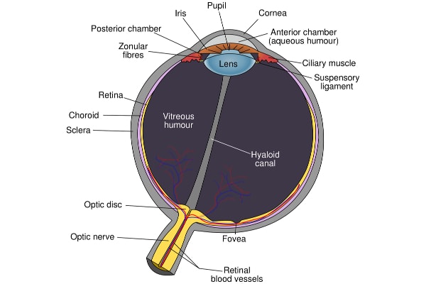
The first location, the uvea, is actually a collection of three structures within the eye. It includes the iris (i.e. the colored portion of the eye), the ciliary body (which lies just behind the iris), and the choroid (i.e. the middle layer of the back of the eye).
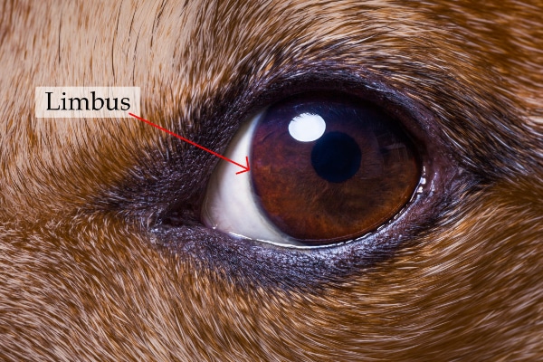
And the other location, the limbus, is the junction between the cornea (the clear front part of the eye) and the sclera (the white, outermost part of the eye).
Uveal melanoma
While relatively uncommon overall, uveal melanomas are the most common primary intraocular tumor in dogs. The majority of uveal melanomas come from the iris or ciliary body, but some can develop from the choroid. Thankfully, over 80% of uveal melanomas are benign.

The remaining 20%, which are malignant, tend to be locally invasive. This means that they can grow into other parts of the eye or, in rare cases, into the brain. However, occasionally, a malignant melanoma can also spread to other parts of the body like the lungs, liver, or regional lymph nodes.
Uveal melanomas are typically round, raised, brown or black pigmented masses. Iris melanomas are usually firmly attached to the iris and can often distort the size and shape of the iris. Alternatively, you might see a mass peeking through the pupil if it originated from the ciliary body. However, since the choroid is in the back of the eye, it wouldn’t be possible to see a choroidal melanoma just by looking at your dog’s eye.
While dogs can develop a uveal melanoma at any age, they are more common in middle-aged to older dogs with an average age of nine years.
Limbal melanoma
Also known as epibulbar melanomas, limbal melanomas originate from the pigmented cells within the limbus. Since the limbus is on the outside of the eye—between the cornea and sclera—limbal melanomas are usually much easier to notice than uveal melanomas.

Most limbal melanomas are rounded, smooth, well-demarcated, and brown or black-colored growths. However, it is possible for a dog to develop an amelanotic melanoma (i.e. one that lacks obvious pigment). Limbal melanomas tend to appear on the side of the eye that is furthest away from the nose.
Melanomas arising from the limbus tend to grow very slowly and are almost always benign tumors. However, if they continue to grow, limbal melanomas can harm the cornea and the sclera. And in extreme cases, these tumors can affect so much of the eye that they lead to vision impairment.
Overall, limbal melanomas are more common in young dogs and senior dogs. However, they can occur at any age.
What causes eye melanomas in dogs?
Like many other types of tumors, the underlying cause of uveal and limbal melanomas in dogs is unknown. But researchers believe genetics may play a role. It is possible that dogs with greater skin pigmentation may have a higher risk of ocular melanomas. The breeds most likely to develop eye melanoma are:
- Labrador Retrievers
- Golden Retrievers
- German Shepherd dogs
- Schnauzers
- Cocker Spaniels
Current research does not support any environmental factors as a cause for ocular melanoma. So, for now, it seems unlikely that UV-ray damage from sunlight is a factor. However, we do know that dogs who spend a lot of time outdoors may be more likely to develop cataracts in dogs. Thus, it may still be a good idea to break out the Doggles (or other dog eye UV protection) when you go hiking or boating with your canine companion.

What are the symptoms of dog eye melanoma?
Based on the fact that limbal and uveal melanoma occur in different locations on the eye, it makes sense that they would have different symptoms.
Uveal melanoma symptoms
Since uveal melanomas grow within the eye, they are harder to notice in the early stages. This is especially the case for ones that arise from the ciliary body or choroid because those structures are hidden behind the iris and in the back of the eye. But even iris melanomas can be easy to miss at first because the iris is already highly pigmented.
In the early stages of uveal melanoma, the dog may be asymptomatic. And you may not know anything is wrong until the tumor is large enough for the vet to visualize it on a physical examination or it starts causing problems.
Eventually, with uveal melanomas that develop in the front of the eye, you may start to see changes in the appearance of the eye. For example, you might notice that the iris has changed shape (i.e. dyscoria). Or you may see blood in the front chamber of the eye (i.e. hyphema).
Additionally, intraocular tumors like uveal melanoma can cause increased pressure within the eye (i.e. secondary glaucoma in dogs). The discomfort associated with glaucoma can cause headshaking, squinting, eye-rubbing, and whining.
Limbal melanoma
Unlike uveal melanomas, limbal melanomas can easily be seen with the naked eye (no pun intended). This makes it easier to detect them in the early stages. Dogs are often asymptomatic at first. However, sometimes limbal melanomas can grow large enough that it becomes difficult for the dog to close the eyelids.
Also, if other surrounding eye structures are involved, limbal melanomas may cause excess tear production and redness or inflammation. And while limbal melanomas don’t usually affect intraocular pressures, they can be painful once they infiltrate the cornea.
Symptoms of both melanoma types
Vision loss is possible with both melanoma types. But because only one eye is affected, dogs may compensate with the opposite eye. Thus, they may not show obvious symptoms of blindness.
Other possible side effects from dog eye melanoma include:
- Uveitis (i.e. uveal inflammation)
- Buphthalmos (i.e. a bulging, enlarged eyeball)
- Lens subluxation (i.e. lens partially moving out of place)
- Retinal detachment
How is ocular melanoma diagnosed?
If you notice a dark spot on your dog’s limbus or iris, or if you see other eye symptoms, schedule a veterinary visit. Your vet can help you figure out what the next steps should be.

It isn’t possible to determine if a melanoma is benign or malignant based on appearance alone. And there are several other conditions (e.g. iridial cysts and iris nevus/freckles) that can look like a melanoma. Therefore, if your vet suspects ocular melanoma or finds any ocular pigmented masses bigger than three millimeters wide, he or she may refer you to a veterinary ophthalmologist for a more in-depth evaluation.
Depending on what the ophthalmologist sees on the ophthalmic exam, he or she may recommend additional testing such as gonioscopy (i.e. looking at the area where the iris and cornea meet), ultrasound, or magnetic resonance imaging (MRI). These tests allow the ophthalmologist to further evaluate the tumor and the eye itself.
Since many uveal melanomas and almost all limbal melanomas are benign, the vet or veterinary ophthalmologist may not recommend diagnostic staging to look for signs of metastatic disease at first. However, staging may be a good idea if the tumor type is unknown or if your dog has any signs of skin or oral cancer. Typically staging involves bloodwork, urinalysis, X-rays or ultrasound, and/or fine needle aspiration of nearby lymph nodes.
Additionally, if the veterinarian or veterinary ophthalmologist removes the mass (or the entire eye) he or she can submit it for biopsy. This is the best way to reach a definitive diagnosis for a tumor of the eye in dogs.
What is the treatment for dog eye melanoma?
After completing the diagnostics, the vet or veterinary ophthalmologist will discuss the treatment options. Dog eye melanoma treatment will vary depending on the type of melanoma and how it is affecting the eye.
Monitoring the tumor
In the early stages of uveal melanoma, or with small, slow-growing limbal melanoma, your veterinarian might choose to monitor the tumor at first. He or she will probably have you check in every one to three months initially. And the vet may take pictures of the eye each time for comparison.
But if the mass is large, painful, or growing rapidly, your vet will probably recommend considering treatment.
Surgical removal or laser therapy
Surgical removal is an option for some tumors involving the limbus or iris. A veterinary ophthalmologist will usually perform this surgery. Alternatively, diode laser treatment for iris melanoma or ciliary body melanoma has a high success rate. In some cases, especially with limbal melanoma, the veterinary ophthalmologist may consider combining debulking of the tumor (i.e. removing as much as possible) and diode laser therapy to decrease the chances of reoccurrence.
Enucleation
If the entire tumor cannot be surgically removed, is growing rapidly, or is causing glaucoma, pain, or other complications, your veterinarian may talk to you about removing the eye entirely (i.e. enucleation). This sounds like a scary option. But keep in mind that many dogs adapt quickly and easily to having one eye.

Enucleation is also the fastest way to alleviate pain and symptoms from invasive ocular melanoma. It does not require long-term use of eye medications or following up with the vet every few months. And submitting the entire eye for analysis is the easiest way to reach a definitive diagnosis. Enucleation is also the most cost-effective treatment option.
Additional options
There are also a few other treatments that may help in some situations.
- Cryotherapy may be an option for limbal melanoma treatment, but it only stops cancer cells from growing. It does not shrink the tumor.
- The melanoma vaccine for dogs (usually available through veterinary oncologists) might not help with treating eye melanomas. But there is some data to suggest that it can help with the metastatic disease caused by uveal melanoma.
- Topical steroidal and non-steroidal eye medications may help relieve painful uveitis.
What is the prognosis for ocular melanoma?
Thankfully, with treatment, the prognosis for dogs with uveal melanoma is good. Most uveal melanomas are benign and the metastatic rate is very low. However, there is still the chance for local invasion and pain if left untreated.
And limbal melanoma is almost always benign and slow growing, so electing to monitor this tumor may be a safe option for your dog. Just keep in mind that any fast-growing mass should be quickly addressed to prevent damage to vision or eye structures.
Work with your vet to keep an “eye” on your dog’s eyes
As we discussed, some dog eye melanomas are easier to see than others. But it certainly doesn’t hurt to get into the habit of looking at your dog’s cornea, sclera, iris, and other visible eye structures on a regular basis. (To learn more about how to perform a weekly dog wellness scan, check out Dr. Buzby’s Tip-to-Tail Health Scan™ course). This routine health scan can be a great way to catch eye problems early on.
Plus, it is a good idea to take your puppy or adult dog to the vet for yearly wellness exams. And you should ideally bring your senior dog to the vet twice yearly for a checkup. These visits allow the vet to examine your dog’s eyes with the ophthalmoscope (and check the rest of your dog out too). Since some ocular melanomas aren’t visible to the naked eye, routine vet checks are a great way to detect melanoma before it starts causing problems.
Finally, if you or your vet find a potential eye melanoma, don’t lose hope. Follow your vet’s instructions (including pursuing a referral to a veterinary ophthalmologist if needed) and take it one step at a time. Chances are good that with careful monitoring or treatment, the melanoma won’t cause any long-term problems.
Did your dog have an eye melanoma?
Please share his or her story below.


My dog Penny has just been diagnosed with malignant melanoma. We had an enucleation done a week ago. Not sure what next steps are. Local vet suggested scans to see if there if it has spread to lungs or other areas. This has all happened very fast and is very overwhelming.
Hi Natalie,
I am sorry Penny has been through so much over the past few weeks. I agree you may need to do some more testing to ensure this cancer has not moved to other areas of the body. Don’t forget you can ask for a referral to an oncologist if needed. Hoping your girl is healing well and praying for a full recovery. Wishing you all the best for brighter days ahead.
Hi everyone, so I’m freaking out! Took my 6 yr old English retriever to our local vet Friday morning after noticing that she had a haze covering her eye and a couple of hours later she had bleeding around her pupil Thursday evening.
Local vet said she had glaucoma and said her pressure in her left (bad) eye was over 70. Sent us home with drops and scheduled a follow up in a week.
Once we got home I just had an uneasy feeling, mostly because her eye was literally changing in a matter of hours. I then called UVH Shreveport LA to see if they could get her in. They had me scheduled to come in at 4pm. They got all the local veterinary information so they could get the records and it was literally 10 minutes later that the vet from UVH called and said load her up NOW! With an eye pressure 50 it’s considered an emergency.
After a hour drive and getting her out of the car she was already completely blind 😞. They had already called in an Ophthalmologist who examined her immediately.
All we know is she definitely has a tumor behind her eye with secondary glaucoma and he felt like it’s uveal melanoma. Also, yes she is now blind. They are removing her eye in the morning and we’re praying that it is not cancer.
However, I know you should stay off google, but I didn’t and now I’m more confused than ever.
Has anyone else been through something like this that happened so quickly?
Dear Cheryl,
My heart goes out to you as you try to find some answers for your sweet girl. I am so sorry she endured this painful condition and has lost her sight. I hope the surgery went smoothly and pray you received favorable results from the pathology report. Feel free to leave an update and let us know how she is doing. Hoping all is well and your pup is adjusting to her new situation. Bless you both.
I just got back from the vet with my senior dog. I took him regarding a small spot I noticed on his eye the last month. The vet checked him and said it could possibly be melanoma but there is no way to tell for sure until he starts to show symptoms. He said to just bring him back if I notice it getting larger or if he is pain. I am just curious as to if I should get a second opinion or not in case there is something else I should be doing for prevention or any other care in the mean time. Please and thank you for your wonderful and very helpful information! I really did get a better understanding on what melanoma is and the types. I feel a lot better after reading your article!
Hi Elizabeth,
I understand your concern for your pup with this worrisome spot on his eye. It is always ok to get a second opinion or search for more information. If you have unanswered questions or lingering concerns, I highly recommend you ask about a referral/consultation with a specialist. They can give you all the details about possible causes, testing, and treatment. Hoping all is well and praying for a positive outcome. Wishing you and your sweet boy all the best.
Hello,
My 10 yr old mini schnauzer had a small mass on the outer corner of his eyelid for years. In the last 6-8 months it has doubled in size. Our vet removed it and it came back as a malignant melenoma. Vet said since more of the eyelid was not removed due to mot being possible that it would likely grow back. Should we take further action at this time or keep any eye on it and see what happens?
Thank you!.
Hi Lindsay,
I am sorry your senior guy has received this difficult diagnosis. Without playing a personal role in your dog’s medical care, it is hard to make specific recommendations. I encourage you to consider scheduling a consultation with a veterinary oncologist. They can give you all the treatment options and discuss the prognosis associated with each. Your vet should be able to make a referral if needed. Hoping you can get some answers and find the best way forward. Bless you and your sweet boy.
Our golden, Denver, is just 2 years old and we found out yesterday he has a uveal melanoma. The ophthalmic surgeon recommends enucleation , but said we could wait a month and determine how fast growing it is. We are trying to find a doctor that might do the laser procedure to remove most of the melanoma. My question is if they will be able to biopsy that part and determine if the growth is malignant. He said it would probably continue to grow. Do you determine this approach to risky? We hate for Denver to lose an eye at just 2 years old as goldens have a risk of other eye disease which could affect his other eye at some point. Thank you from Austin Texas.
Hi Brenda,
I am sorry Denver has received this difficult diagnosis. I understand your concern with enucleation, but in my experience, most dogs do very well with only one eye, and this could be the only way to ensure the cancer is completely resolved. They should be able to submit the eye tissue for pathology to review and give you an assessment of malignancy or if it seems benign. Ultimately, in cases like this, I defer to the recommendations of the specialists. If the ophthalmic surgeon wants to remove the eye, then I would probably remove the eye. Wishing Denver a successful surgery and a smooth recovery. Please keep us updated on how he is doing!
Thank you for your article!
We just recently had an enucleation done on our dogs eye and after reading this, we feel better about the speed in which we decided to have the operation done (approx 4 weeks after we noticed her Iris change, hyphema, & saw the ophthalmologist). Pathology hasn’t come back yet, but X-rays of the lungs didn’t show any spotting and the vet said they didn’t see any discoloration in the ocular cavity.
What sort of questions should I ask when pathology comes back?
Hi Robert,
I am glad the surgery went well and think it is great you are staying on top of things. The pathology results themselves may answer all your questions. Many times, they will contain what next steps are recommended by the pathologist. But the main thing to find out is if any further treatment needs to be done and if there will be any follow up appointments or things to monitor for in the future. Hoping you receive good news. Feel free to keep us updated as you continue your journey.
I’m at the vet with my 10 yr old ACD mix Smokey. She has bleeding inside the eye that I first noticed as one eye being darker brown/red than the other. The vet has detected a mass they think could be uveal melanoma and is suggesting enucleation. We’ll see where it goes and try to update this post when we know. Thanks for posting this information, very helpful!
Hi Dan,
I’m so sorry to hear that Smokey may have a uveal melanoma. I hope all goes well with the surgery and that you get good news on the pathology report. Please do keep us updated on how things go. Thinking about you during this stressful time of waiting. ❤️