Glaucoma in dogs (aka high pressure within the eye) can be a painful and sight-stealing condition. Integrative veterinarian Dr. Julie Buzby explains the causes, symptoms, diagnosis, and treatment of glaucoma to help devoted dog parents feel more prepared should their dog develop glaucoma.
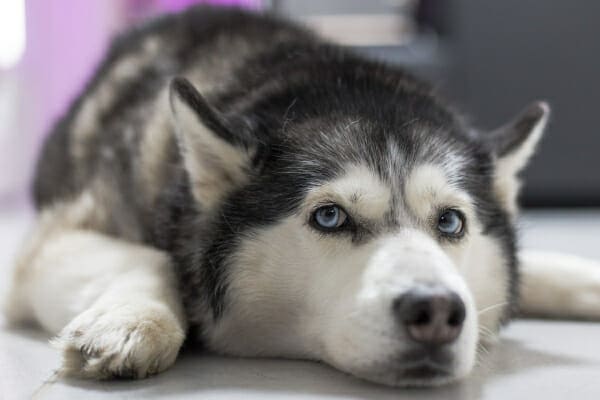
As a veterinarian, I often say “time is of the essence” when it comes to eye problems in dogs, and glaucoma is no exception. Once the ball starts rolling, glaucomatous eyes can deteriorate quickly, leading to consequences such as significant pain and loss of vision.
While glaucoma does tend to progress over time, the good news is that there are a variety of medical and surgical options that can help keep your dog visual and comfortable for as long as possible. And thankfully, even if your dog goes blind or needs to have the eye removed due to glaucoma, most non-visual dogs adjust well and continue to lead happy lives.
Let’s take a closer look at canine glaucoma.
What is glaucoma in dogs?
Glaucoma in dogs is a medical condition that causes increased pressure within the eye (aka intraocular pressure or IOP). It may affect one or both eyes in dogs. Unfortunately, glaucoma can cause severe damage to the affected eye(s).
If nerve signals and/or blood flow to the eye’s internal structures are compromised, it can result in permanent blindness due to the death of retinal cells. (The retina is a thin layer of specialized sensory cells that line the inside of the back of the eye, responsible for capturing reflected light to send the image information to the brain via the optic nerve).
Additionally, the excessive pressure within the eyeball is extremely painful. Over time, chronic pain from a glaucomatous eye can significantly decrease a dog’s quality of life.
What causes glaucoma in dogs?
Glaucoma in dogs is caused by a change in intraocular pressure (IOP).
In glaucoma, the IOP increases because there is a decrease in the drainage of aqueous humor. The aqueous humor is the clear fluid that fills the portion of the eye in front of the lens (a clear, round disc behind the iris that focuses light onto the retina) and is very important for overall eye health.
Aqueous humor keeps the lens clean and carries nutrients from the blood to various eye structures. Once nutrients have been used up, it makes sense that the older aqueous humor must be replaced by newer fluid.
In a normal dog, there is a delicate balance between aqueous humor production and drainage, which helps keep the IOP at a normal level. Aqueous humor is produced by the ciliary body, which is the ring-shaped muscular tissue that controls the size of the pupil.
However, IOP will increase if aqueous humor cannot drain out of the eye via the iridocorneal angle (ICA), the angle formed between the cornea (the clear, outer layer on the front surface of the eye) and iris (the colored part of the eye that controls how much light moves through the pupil).
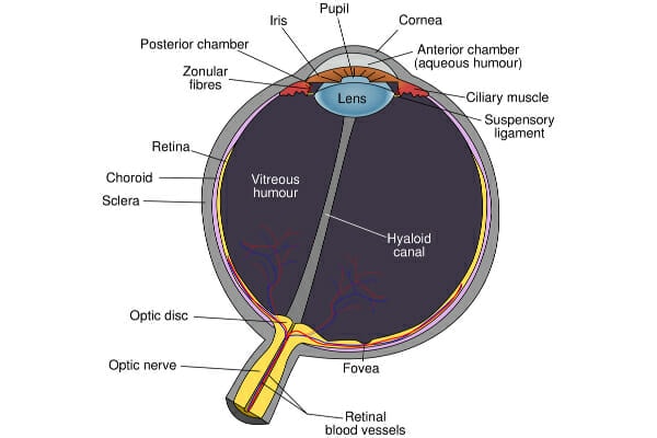
The ICA determines how quickly aqueous humor flows out of the eye. IOP increases because the ciliary body is still producing new aqueous humor even though it’s not properly draining.
Think of it like plumbing—a damaged or clogged drain will cause water to back up in the sink if the faucet is not turned off. So, if aqueous humor cannot flow out of the eye, more of it will accumulate inside the eye because it has nowhere else to go. And more fluid crammed into the same-sized space in the eye means the intraocular pressure will increase.
Types of glaucoma
Glaucoma is divided into two categories: primary and secondary. Primary glaucoma is typically inherited and is more common in purebred dogs. On the other hand, secondary glaucoma means that the increased IOPs are due to some other factor besides genetics.
Primary glaucoma almost always affects both eyes, whereas secondary glaucoma may only affect one eye.
Primary glaucoma
Dogs with primary glaucoma have typically inherited one or multiple gene mutations that lead to malformations of the iridocorneal angle (like closed-angle glaucoma, where a smaller ICA affects drainage flow) or of the pectinate ligaments, which are a network of fibers at the ICA. The spaces in between these fibers act like drains for aqueous humor.
In goniodysgenesis, for example, the fibers of the pectinate ligaments are replaced by sheets of tissue. Think about this like trying to drain water through a colander with tiny holes instead of big holes.
In patients with open-angle glaucoma, the ICA is normal-sized, but cartilage building blocks called glycosaminoglycans (GAGs) accumulate in the network of pectinate ligaments, causing a physical obstruction. This is sort of like how the drain in your shower may become slower and slower as hair accumulates in it.
Breeds predisposed to primary glaucoma
There are specific dog breeds that are more likely to develop certain gene mutations causing primary glaucoma. Genetic testing for these mutations is available for some breeds.
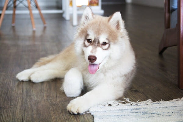
Dog breeds that are more prone to developing closed-angle (aka narrow-angle) glaucoma include the following:
- Akitas
- Basset Hounds
- Chinese Shar Peis
- Chow Chows
- Cocker Spaniels
- English Springer Spaniels
- Flat-Coated Retrievers
- Poodles
- Samoyeds
- Shiba Inus
- Shih Tzus
- Siberian Huskies
Open-angle glaucoma occurs more commonly in:
- Beagles
- Norwegian Elkhounds
- Petit Basset Griffon Vendéens
For both primary and secondary glaucoma, gender does not appear to influence the risk of developing glaucoma. The age of onset is variable, but for primary glaucoma, middle-aged dogs are the most affected.
Although rare, primary glaucoma in puppies does occasionally occur. This congenital condition is usually diagnosed between three and six months of age. It can affect one or both eyes and sometimes occurs in conjunction with other eye abnormalities.
Secondary glaucoma
In secondary glaucoma, there are multiple eye issues that can lead to increased IOPs. Ocular trauma and lens luxations are among the most common causes. Uveitis in dogs (inflammation of the uvea, which is composed of the iris, ciliary body, and choroid) is also a contributing factor. For example, cataracts in dogs can cause uveitis, which may then lead to glaucoma.
Additionally, ocular cancer can affect aqueous humor drainage from the eye.
Sudden onset glaucoma in dogs
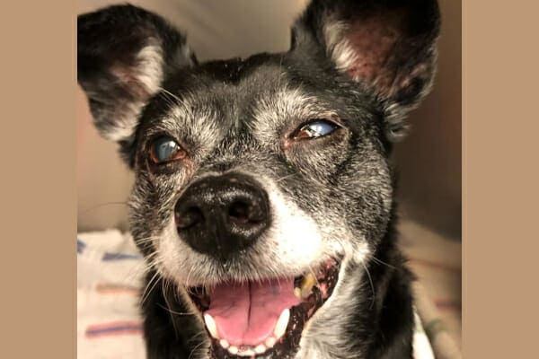
Health issues that can cause hypertension in dogs (elevated blood pressure) may also increase IOP. Some of these include heart disease in dogs and Cushing’s disease in dogs. In most cases of sudden-onset glaucoma in dogs (aka acute glaucoma in dogs), there is an underlying problem of some sort that leads to secondary glaucoma.
Secondary glaucoma can affect most dog breeds because the contributing factors are numerous. However, Boston Terriers and Fox Terriers may be more at risk of developing lens luxations that can progress to glaucoma.
What are the symptoms of glaucoma?
The symptoms of glaucoma in dogs can be similar to other eye conditions, so it’s very important to have your dog evaluated right away if you notice changes in eye appearance or vision.
Early signs
With sudden onset glaucoma, you may see some of the following symptoms:
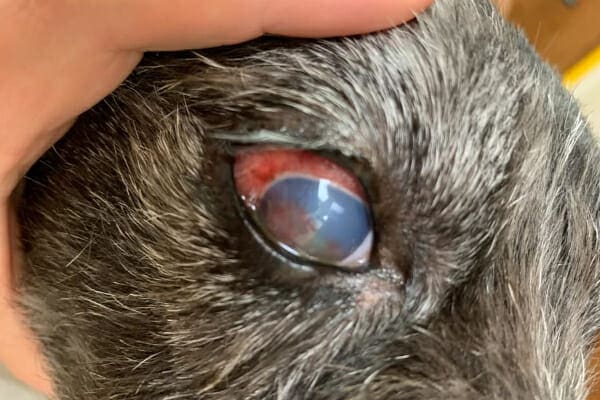
- Corneal edema: A bluish-white discoloration of the cornea caused by fluid accumulation
- Episcleral injection: Blood vessels that have become enlarged and more noticeable across the sclera (the “white of the eye”)
- Conjunctival hyperemia: Redness of the normally pink tissues around the eye
- Mydriasis: Dilated pupil
- Eye discharge: Usually watery and clear
- Blepharospasm: Excessive eyelid blinking and squinting
- Pain: Rubbing or pawing at the eyes, attitude changes, or other signs your dog is in pain
- Blindness: Bumping into things or seeming hesitant to move if both eyes are blind, but this may not be noticeable if only one eye is affected
Seek veterinary care immediately for signs of glaucoma
If you notice any of these signs, please make an appointment with your veterinarian promptly. Especially with sudden-onset glaucoma, time is of the essence. High IOPs can damage the retina, sometimes permanently. So, the sooner the vet is able to diagnose glaucoma and reduce the IOP, the better the chance of preserving the dog’s vision.
Later signs
If the IOP remains elevated, a dog may progress to chronic glaucoma. Eventually, during end-stage glaucoma, the eye may appear to be larger than normal (buphthalmic), and the dog may be completely blind in that eye. If the dog only has glaucoma in one eye, signs of vision loss may not always be very obvious because the “good” eye can compensate.
Is glaucoma in dogs painful?
Glaucoma in dogs is quite painful. If you think about having too much pressure in your eyes, you can imagine how uncomfortable that would be. That’s why dogs with glaucoma may paw at or rub their faces. They just don’t know what to do to feel better!
If you ever suspect your dog may have glaucoma, take them to the vet right away so they can get some relief. Even the early stages of the disease cause significant pain.
How will the vet diagnose my dog with glaucoma?
In order to diagnose your dog with glaucoma, your veterinarian will perform a complete physical exam to look for signs of underlying health issues. Plus, your vet will thoroughly examine your dog’s eyes.
Measuring eye pressure
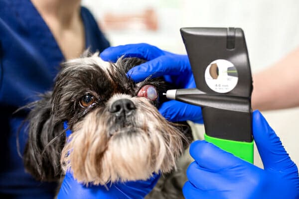
Many veterinary clinics have a special tool called a tonometer. The vet can use the tonometer to measure a dog’s IOPs by briefly contacting the surface of the eye. Depending on the type of tonometer, your vet may place a drop of numbing solution in each eye to prevent discomfort during the examination.
Intraocular pressures are measured in millimeters of mercury or mmHg (just like atmospheric pressure). A normal range of values should be between 10 and 20 mmHg, but can read as high as 24 mmHg with certain tonometer types. The vet will generally classify readings higher than 25 mmHg as abnormal. He or she may also suspect an eye issue if there is a difference of more than 8 mmHg between the two eyes.
It is important to note that one elevated value may not be enough to definitively diagnose glaucoma. The vet likely will repeat the tonometry and only suspect glaucoma when the IOPs are consistently high.
Additional testing
Genetic tests for the mutations that lead to primary glaucoma are available for Shar Peis, Beagles, and Petit Basset Griffon Vendéens. Your vet may also recommend testing that looks for signs of other illnesses.
For example, as mentioned previously, conditions that cause high blood pressure, like heart disease, kidney disease, and various endocrine disorders (i.e., Cushing’s disease or hypothyroidism in dogs), can lead to secondary glaucoma. It may be a good idea to run blood tests for dogs who develop glaucoma to help rule out those conditions.
In some cases, your vet may refer you to a veterinary ophthalmologist for further evaluation. These eye specialists have other tools for evaluating glaucomatous eyes. For example, they can use a unique lens to perform gonioscopy, which is the evaluation of the iridocorneal angle. The overall appearance of the ICA can distinguish between secondary and primary glaucoma.
They are also capable of performing some surgical procedures for glaucoma treatment and/or developing a medical management plan for cases of glaucoma that are difficult to control.
How do you treat glaucoma in dogs?
Treating glaucoma in dogs can include various medical or surgical options. Once your vet diagnoses your dog with glaucoma, he or she will discuss the different treatments available.
Overall, most canines with glaucoma respond better to surgical intervention in the long run. This is especially true for dogs with primary glaucoma, lens luxations, or ocular tumors. However, glaucoma surgery can be expensive, and it isn’t the right option for all dogs, so medical management may also be reasonable.
Medications for glaucoma in dogs
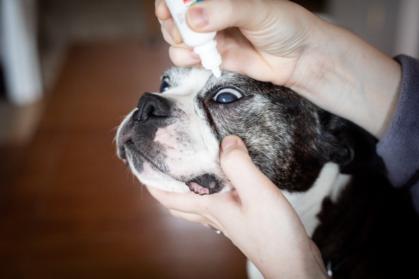
Sometimes the veterinarian may recommend starting with medical therapy such as glaucoma eye drops or oral medications. These can include:
Prostaglandin analogues
As mentioned earlier, acute glaucoma may damage the retina, leading to blindness. So, in an emergency situation, severely elevated IOPs may warrant the use of a medication that rapidly lowers pressures in order to ideally preserve a dog’s sight.
One class of topical eye medications, called prostaglandin analogues, can help with this. These medications, such as latanoprost, decrease resistance to aqueous humor outflow.
It is worth noting that prostaglandin analogues are only intended for short-term use. Additionally, they may cause temporary blindness because they constrict the pupils.
Carbonic anhydrase inhibitors
The most common type of glaucoma medication for long-term use is dorzolamide, which is a carbonic anhydrase inhibitor (CAI) drug. Inhibition of the enzyme carbonic anhydrase will decrease aqueous humor production. As a result, the IOP becomes lower.
Beta blockers
Your vet may also prescribe beta-blocker medications like timolol because they also decrease the production of aqueous humor by the ciliary body. However, your vet must use caution when using this drug in dogs with airway problems or heart disease.
Additional medications
If your dog develops secondary glaucoma due to uveitis, your vet may recommend topical steroid therapy.
Additionally, all dogs with acute onset glaucoma and some with chronic glaucoma can benefit from oral pain medications. It is important to keep in mind, though, that in dogs with severe glaucoma, oral pain medications aren’t typically sufficient to relieve the pain as long as the IOP is also high. Thus, it is important to manage IOP and manage pain.
In the event that medical management fails or is not an option, surgical treatment will be necessary. The surgical technique that the vet or veterinary ophthalmologist selects will depend in part on whether or not your dog still has vision in the affected eye.
Surgery for glaucoma in dogs
Surgery may be an option for the treatment of glaucoma in some dogs. If the dog can still see, the goal of surgery is to manage IOP and preserve vision for as long as possible.
One option is a procedure called cyclophotocoagulation. In it, the veterinary ophthalmologist uses a surgical laser to destroy a part of the ciliary body. Doing so causes a decrease in aqueous humor production.
Sometimes, the veterinary ophthalmologist can implant a shunt that diverts aqueous humor around a diseased ICA so that it can leave the eye. This surgery is called gonioimplantation. The ophthalmologist may choose to combine it with cyclophotocoagulation in more difficult cases.
Finally, if the dog has a concurrent lens luxation, the veterinary ophthalmologist can surgically remove the lens. This should improve the secondary glaucoma.
Surgery for blind dogs
Unfortunately, sometimes glaucoma causes permanent blindness. In that case, the focus switches to keeping the dog as comfortable and pain-free as possible.
Enucleation
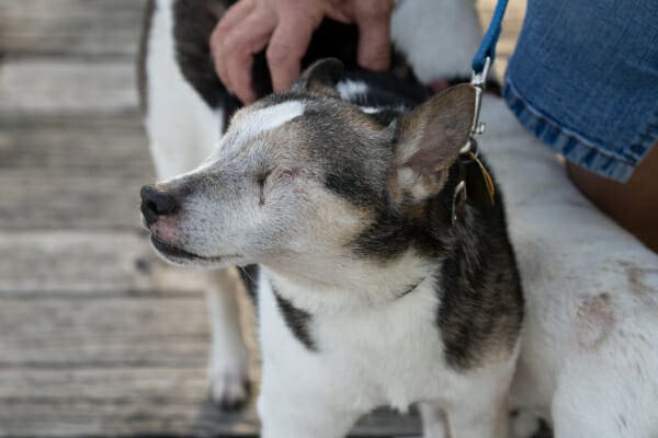
It may sound scary, but sometimes enucleation surgery is the best option for dogs with painful, blind eyes who are in end-stage glaucoma. For this procedure, the vet or veterinary ophthalmologist will surgically remove the entire glaucomatous eyeball.
From a cosmetic standpoint, your canine companion will look like he or she is permanently winking at you.
Although it may be difficult to think about your dog losing an eye, the great thing about enucleation is that it provides immediate relief from pain and discomfort. If the eye is not visual and will not regain vision, there is no benefit to keeping it.
Dog parents often report that their dog starts acting like a puppy again after surgery because he or she is not in constant pain from the eye anymore.
Additionally, enucleation is typically less expensive than the other surgical procedures and does not require specialized equipment. This often means your family veterinarian can perform the enucleation surgery.
Intraocular prosthesis
If you would like to avoid enucleation surgery, your veterinary ophthalmologist can discuss a few other options.
One option involves the veterinary ophthalmologist removing the eye’s internal structures and replacing them with an ocular prosthesis for dogs. Some dog parents find this type of surgery more cosmetic than an enucleation. Plus, it does still provide pain relief.
However, this is not a good treatment for dogs with ocular cancer, dry eye in dogs, chronic inflammation, or chronic corneal disease.
Gentamicin injection
Another procedure involves injecting gentamicin, an antibiotic, into the eye in order to destroy the ciliary body. This is only useful for cases of chronic glaucoma and has a 65-80% success rate. Ultimately, you may still need to consider enucleation if this procedure fails.
Can glaucoma in dogs be cured?
Unfortunately, glaucoma in dogs is not a curable disease. Medical therapies and surgery can help prolong progression and manage pain. But a dog with glaucoma will almost certainly eventually become non-visual.
Are there home remedies for glaucoma in dogs?
There aren’t really any effective home remedies for glaucoma in dogs, but that doesn’t mean that what you do at home doesn’t matter.
Administering medications and following post-op instructions
For example, it is important to administer glaucoma eye drops or oral medication according to your veterinarian’s instructions. It is also critical to follow any post-op instructions should your dog have glaucoma surgery. Good nursing care and appropriate medication use can go far in helping your dog be as comfortable as possible.
Monitoring your dog’s eyes closely
Additionally, since you see your dog’s eyes every day and your vet does not, you have the important job of watching for subtle changes that may indicate the IOP is rising again, or the dog is developing another problem.
If the eyes start to look different (red, cloudy, etc.), or your dog starts to squint or act painful, contact your vet immediately. The sooner the vet can (hopefully) get the IOP under control, the better the chance the eye will remain visual.
Helping your dog adjust to being blind
If your dog is blind, there are also some things you can do to help him or her adjust. You can help care for your blind dog by:
- Keeping furniture in the same location
- Always having the food and water in the same place
- Placing a rug or a different textured surface at the doorways or stairs to help the dog know what is coming up
- Frequently talking to your dog so he or she knows where you are and feels reassured in new situations
Also, although I didn’t design Dr. Buzby’s ToeGrips® dog nail grips with blind dogs in mind, I have had numerous people testify to how much they helped their blind dogs navigate the world with confidence.
Being unsure of your footing can be unnerving for any dog, but especially for a blind dog. So, it makes sense that the added traction of ToeGrips for blind dogs can be very reassuring.
What is the outlook for dogs with glaucoma?
When thinking about the prognosis for dogs with glaucoma, it is important to differentiate between the prognosis for remaining visual and the prognosis for remaining comfortable.
Prognosis for vision: Guarded
Unfortunately, almost all dogs with primary glaucoma will eventually become non-visual. For dogs with secondary glaucoma, the prognosis for retaining sight in the eye depends on the cause. Some causes are more treatable than others.
The good news in this, though, is that many dogs adapt well to being blind. And they can still go on to live happy, full, and long lives.
Prognosis for comfort: Good with appropriate measures
The prognosis for comfort, however, is much better with appropriate treatment and a willingness to pursue enucleation if needed. For a period of time, medical and/or surgical management may be able to keep the IOP, and therefore pain, under control.
Once your dog’s pain isn’t well-managed, it is time to pursue an enucleation or similar solution. Removing that painful eye can greatly improve your dog’s comfort and quality of life long-term. He or she won’t need daily eye drops and will be in a lot less pain. (Remember, though, end-stage glaucoma is extremely painful. So, on the flip side, the prognosis for comfort long term is poor without enucleation.)
When to put down a dog with glaucoma
The good news is that just because your dog has glaucoma, that doesn’t mean you should put him or her down right away. As you can see from the long list of treatment options shared above, your vet has a lot of tools to help manage glaucoma.
And even if your dog becomes blind, that doesn’t mean that his or her life is over. Blind dogs can still have a wonderful quality of life with a dedicated owner and good pain control.
End stage glaucoma in dogs
When you do get to the point where your dog isn’t coping well with being blind, or you can no longer get the pain under control and enucleation isn’t an option, then it may be time to have a conversation with your vet about whether preparing for your dog’s euthanasia is the kindest option.
Partner with your vet after a canine glaucoma diagnosis
If you suspect your dog might have glaucoma (or another eye problem), seek veterinary care immediately. Glaucoma treatment options are becoming more widely available and reasonable. And prompt medical and/or surgical treatment can help your dog to be comfortable and visual for as long as possible.
After the diagnosis, take steps to control your dog’s IOP with medications or surgery, be willing to do what it takes to minimize his or her pain (even if that means an enucleation), and stay in close communication with your vet.
Hopefully, you and your dog will be able to enjoy many more wonderful years together after a glaucoma diagnosis and make many, many more lasting memories.
Has your dear dog been diagnosed with glaucoma?
Please share his or her story in the comments.
This post was originally published on February 7, 2022 and updated on May 18, 2026.

I am so sorry for the dogs that have eye problems I did have a Maltese years ago that had retinitis pigmentosa she went blind and we did everything to help her put soft toys on the side of tables so she wouldn’t; hurt herself etc she did well considering but it was hard on us watching our sweet little girl my question is there anything we can do to help with eye health my dog now is a Miki who is 12 his eyesight is good but I notice a very little cloudiness in his eyes but wondering what can do to help eyesight in older dogs either in food or supplements.
Hi Tina,
This is a great question. I am not sure I have any specific recommendations for food or supplements for eye health. There are several available online, but I can’t speak to their effectiveness or safety. My best advice is to have your vet check Miki’s eyes each year at his routine wellness exam. If they notice any problems (dry eye, cataracts, ulcers, etc.) they can act quickly and start treatment right away. Unfortunately, diminished eyesight is a common problem that tends to come with old age. Hoping your boy can avoid any major issues and wishing him a long and happy life.
I have a 12 year old Husky who had cataracts but over a three day period the blue eye developed Glaucoma the vet gave us eye drops and medication for the pressure. After one week the vet determined she was blind in that eye and now wants to remove it. Is there no other options?
Hi Greg,
I am sorry your Husky has lost the vision in her eye and is battling this difficult condition. As the article states, there are a few different medications that can be used to try and maintain normal eye pressures, but once vision is lost, removing the eye is the most common treatment. If the eye is nonvisual, then there is really no benefit to trying to keep the eye in place. But once the affected eye is removed, the pain is gone, and most dogs feel instantly better. I am curious to know what your concern is with the enucleation surgery. Is it the anesthesia? Or how she will look afterward? Don’t hesitate to discuss any lingering issues with your vet. You deserve to feel confident and be at peace with the choices being made. Wishing you all the best. Feel free to leave an update if you have a chance.