Cloudy eyes in dogs can happen for a variety of reasons—some are more concerning than others. To help you sort through possibilities, integrative veterinarian Dr. Julie Buzby explains the symptoms, diagnosis, and treatment for several common causes of cloudy eyes.
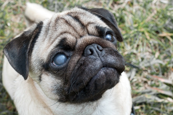
You’ve probably heard the expression that the eyes are the windows to the soul. But did you know that looking into your senior dog’s eyes might also tell you something about his or her health? For example, cloudy eyes in dogs can point to a variety of eye problems.
In some cases, the cloudiness builds gradually, and in others it appears suddenly. Cloudy eyes may sometimes require emergency treatment, whereas other times the situation is less dire. Being aware of some of the causes of cloudy eyes in dogs can help you know what to do if you notice a change in the appearance of your dog’s eyes.
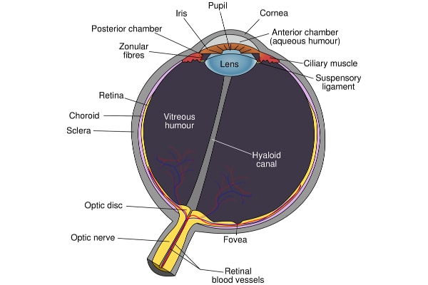
The anatomy of a cloudy eye in dogs
Normal, healthy eyes tend to be clear in the front. This gives you a “window” you can look through to see the iris, pupil, and other structures within the eye. Even the lens, the disc-like structure behind the iris, should be crystal clear.
In fact, the outer and inner structures should be so transparent that you can see all the way back to the shiny back part of the eye (the tapetum lucidum). This is the part of the eye that creates the “green eye glow” when you shine a light toward your dog in the dark.
What are cloudy eyes in dogs?
If your dog’s eye appears grayish, rather than showing their clear black pupil, this may indicate a change or problem in one of the parts of the eye.
Any opacity or cloudiness affecting a particular ocular structure can change the overall appearance of the eye. And depending on the problem, it may even impact your dog’s ability to see. As one of my ophthalmology professors used to say, “If you can’t see in, the dog probably can’t see out.”
Locations of the cloudiness
Because there are so many different medical problems that can cause cloudiness in a dog’s eye, it is sometimes easiest to classify the causes of cloudiness based on where the opacity is occurring. There are three different segments to consider:
- The cornea—This thin, clear layer is the “windshield” at the front of the eye. It is rich in nerves, which makes it very sensitive to painful stimuli. If the whole cornea becomes cloudy, you cannot see into the eye at all.
- The aqueous humor—This clear fluid surrounds the iris and fills the portion of the eye in front of the lens. Sometimes the aqueous humor can contain red blood cells, white blood cells, protein, or other substances that turn it from clear to cloudy or hazy.
- The lens—This clear disc-like structure is located behind the iris. It is flexible, which allows it to alter the direction of the light as it projects on the retina (the back layer of the eye, which is responsible for converting the light into a nerve impulse and sending it to the brain via the optic nerve). If the lens is cloudy, you can still see clearly through the cornea and aqueous humor. But the area behind the pupil and iris appears hazy or opaque.
Most common causes of cloudy eyes in dogs
Now that you have a little background in eye anatomy and where cloudiness can occur, it may be easier to understand some of the reasons for cloudy eyes in dogs. Next, we’ll break down seven different ocular disorders into four separate sections:
- The cloudy cornea
- Cloudy cornea and/or aqueous humor
- The cloudy aqueous humor
- The cloudy lens of the eye
Section 1: The Cloudy Cornea
The ultra-thin and normally clear cornea can have a white or bluish coloring to it when it is diseased. This occurs because the cornea must maintain a very strict water balance in order to remain clear. If something upsets that balance, excess fluid accumulates in the cornea and turns it cloudy.
Corneal edema (fluid build-up in the cornea) may occur as a result of corneal damage or a range of corneal diseases. It is a clinical sign and not a specific condition itself. This means the vet will need to investigate further to find the cause of the corneal edema.
#1: Corneal ulcers in dogs
One common cause of corneal edema is corneal ulcers in dogs (defects in the surface of the cornea). Corneal ulcers may occur if a dog scratches his or her eye, gets something foreign or irritating in the eye, or experiences an eye injury. Generally, the dog will have one cloudy eye or a cloudy spot on the eye.
Symptoms of a corneal ulcer in dogs
Initially, the dog’s cornea may still appear clear if the ulcer is very superficial. Or there might be a small cloudy spot of corneal edema surrounding the ulcer. Sometimes, mild ulcers can be tricky to detect with the naked eye. So, you may not know there is an issue unless your dog displays some of the following clinical signs:
- Pawing or rubbing at the eye
- Squinting the eye (blepharospasm)
- Visible red blood vessels on the cornea (neovascularization)
- Excessive tearing or watery eyes (epiphora)
- Signs your dog is in pain when you touch near the eye
Corneal ulcers that affect the deeper layers of the cornea can cause observable defects or holes in the cornea. They also tend to go with more widespread corneal edema and may cause more severe symptoms than superficial ulcers. Without aggressive treatment, corneal ulcers, especially those that are deep, can cause permanent damage and lead to blindness. Thus, it is crucial to make a vet appointment promptly if you suspect your dog has an eye ulcer.
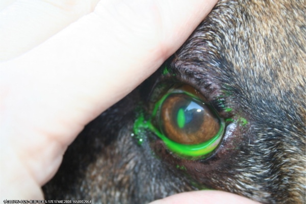
Diagnosis of a corneal ulcer
At the appointment, your veterinarian will use a fluorescein eye stain test to visualize and diagnose the suspected corneal ulcer. This test is typically non-painful and involves applying a drop of fluorescent dye to each eye. If the dog has a corneal ulcer, the dye will stick to the defect in the cornea and glow green when the veterinarian illuminates the eye with a blacklight. If there is no ulcer, the cornea will not take up any stain.
Treatment for a corneal ulcer in dogs
Superficial ulcers are often treated with topical antibiotics and oral medications for pain. However, deeper ulcers may require additional therapies such as drops of serum (the liquid portion of the blood) to promote healing.
#2: Corneal dystrophy in dogs
Alternatively, there are some edematous conditions of the eye that are heritable. Corneal dystrophy is one such condition. Affected dogs will develop corneal edema at some point in their life due to inherited corneal abnormalities.
There are three types of corneal dystrophy:
- Stromal corneal dystrophy—This variety affects the middle layer of the cornea. It is more common in younger dogs, especially Spaniel breeds and Collies.
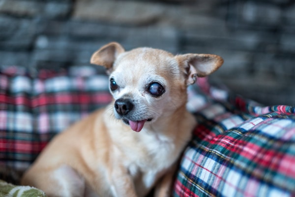
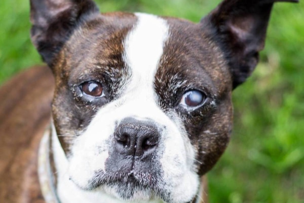
- Endothelial corneal dystrophy—This type affects the inner layer of the cornea and typically shows up in middle-aged to older dogs. Boston Terriers, Dachshunds, and Chihuahuas are all more prone to endothelial dystrophy.
- Epithelial corneal dystrophy—This kind impacts the outer layer of the cornea and affects all dog breeds.
Some canine patients with corneal dystrophy may act normally, other than having cloudy corneas. Others experience discomfort, especially since advanced cases have an increased risk of developing corneal ulcers. For this reason, your vet might still recommend fluorescein testing in order to rule out an active ulcer.
Unfortunately, there is no definitive treatment for corneal dystrophy. However, some veterinary ophthalmologists may recommend topical saline drops to help pull some of the edema away from the cornea. There is also a newer treatment for corneal endothelial dystrophy called Descemet’s Stripping Endothelial Keratoplasty (DSEK), which shows some promise.
#3: Keratoconjunctivitis sicca (dry eye in dogs)
If your dog has a cloudy eye with discharge, he or she may have a condition called keratoconjunctivitis sicca (KCS). Commonly referred to as dry eye in dogs, KCS is any condition that negatively affects tear production in the eye.
Symptoms of KCS in dogs
Dry eye is problematic because tears act like a protective shield for the eye. They lubricate the eye, protecting it from trauma, and can help prevent infection from taking hold. Without adequate tear production, the eyes can become very irritated, red, cloudy, and goopy.
You may see a green or yellow, sticky, mucus-like discharge that builds up over and around the eyes. When it dries, the discharge can look very crusty or like a cloudy film over the eyes. Also, because they are so dry, the surface of the eyes can have a dull appearance. Dry eye can also lead to corneal ulceration in dogs.
Causes of KCS in dogs
The most common cause of KCS is immune-mediated (the immune system attacks the tear gland for unknown reasons). Damaged tear glands, allergies, neurologic disorders, certain medications like sulfa drugs, and endocrine disorders like hypothyroidism in dogs can also cause KCS in dogs. Additionally, there are numerous dog breeds that are more likely to develop KCS, including:
- Boston Terriers
- Cocker Spaniels
- English Bulldogs
- Lhasa Apsos
- Miniature Schnauzers
- Pekingese
- Pugs
- Shih Tzus
- Yorkshire Terriers
Diagnosis of KCS in dogs
If your dog has cloudy eyes and discharge, your regular vet may recommend a series of tests. In addition to the fluorescein eye test for detecting corneal ulcers, the vet may also recommend performing a Schirmer tear test and tonometry.
The Schirmer tear test uses a small piece of metered paper that has a dye-colored line on it. The tip of the paper is slipped beneath the dog’s lower eyelid and left in place for one minute. Any tears the dog produces will cause the colored line to move down the paper. After 60 seconds have elapsed, your vet will look at how far the dyed line has traveled. Anything less than 15 mm is suspicious for dry eye in dogs.
The other test, tonometry, is the measurement of the intraocular pressure or IOP within the eye (more on this in the glaucoma section). This test is important because sometimes canine patients with eye conditions such as glaucoma (high intraocular pressure) will get secondary KCS due to tear gland inflammation.
Treatment for KCS in dogs
Veterinarians can manage most cases of dry eye with a combination of topical lubricants and moisturizers. If the KCS is autoimmune in nature, the vet may prescribe topical immunosuppressants to protect the tear glands from further damage in hopes that the glands might produce tears again.
In special cases, a veterinary ophthalmologist may recommend a surgical procedure for dogs with KCS. It involves redirecting the flow of saliva from one particular salivary gland to the eye. However, this surgery comes with its own set of risks and complications. Your veterinary eye specialist will advise you on what is best for your dog.
Section #2: Cloudy cornea and/or aqueous humor
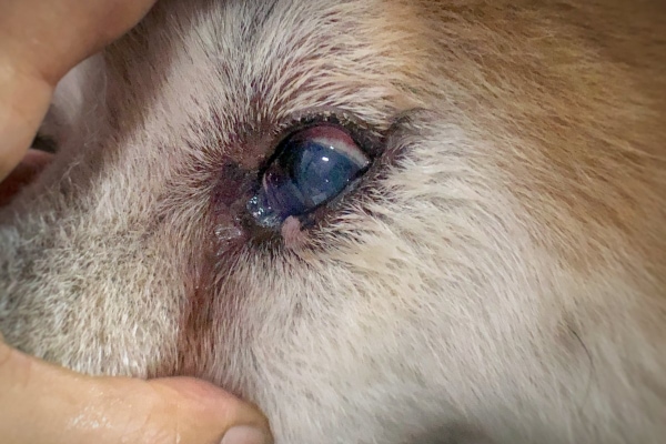
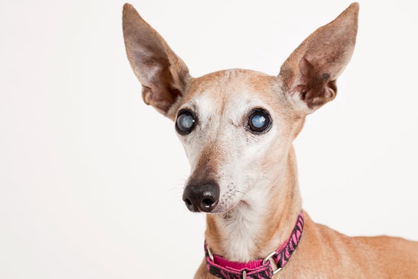
#4: Glaucoma in dogs
As mentioned above, glaucoma in dogs occurs when the pressure within a dog’s eye is elevated. In acute cases, it can cause a dog’s eye to be red, painful, and cloudy. And in more chronic cases, the eye may be cloudy and also enlarged, swollen, or bulging due to the high IOPs.
Cloudiness can occur on the cornea in the form of corneal edema. However, it can also occur within the front (anterior) chamber of the eye. This is called aqueous flare.
Causes of glaucoma in dogs
Glaucoma happens when aqueous humor, which is constantly being produced, cannot properly drain away from the dog’s eye. Normally, aqueous humor is produced at the same rate that it exits the eye via the iridocorneal angle (ICA), a small opening between the cornea and the iris. When something disrupts this balance, the aqueous humor builds up, and the IOP begins to rise.
Glaucoma may either be primary or secondary. Dogs with primary glaucoma have a genetic defect that impairs the ability of the iridocorneal angle (ICA) to drain aqueous humor. But for dogs with secondary glaucoma, the elevated IOPs occur due to some other eye issue (e.g., trauma, lens luxation, hypertension in dogs, ocular cancer, etc.), which impedes the flow of aqueous humor.
Symptoms of glaucoma
Regardless of the type of glaucoma, most of the symptoms are similar:
- Widespread corneal edema, which makes your dog’s eye cloudy and blue
- Episcleral injection (enlarged blood vessels within the white part of the eye, which can make the eye look bloodshot)
- Conjunctival hyperemia (inflammation of the pink tissues surrounding the eyeball)
- Aqueous flare (a beam of light is visible when it passes through the anterior chamber because of the abnormally high protein content of the aqueous humor)
- Blepharospasm (squinting and blinking)
- Eye discharge
Sudden-onset glaucoma is a medical emergency. If you see these signs, please seek an emergency vet visit for your dog. The elevated IOPs can sometimes cause permanent retinal damage and blindness if not quickly addressed.
Diagnosis of glaucoma in dogs
Emergency clinics, as well as some general practice vet clinics, should have a tonometer (a tool to measure IOP). Normal values can be anywhere from 10 to 20 mm Hg, but anything higher than 25 mm Hg may indicate the dog has glaucoma.
To better differentiate between primary and secondary glaucoma, a veterinary ophthalmologist can also perform a specific type of eye exam called gonioscopy to evaluate the ICA. Additionally, genetic tests for glaucoma are available for Shar Peis, Beagles, and other dog breeds that are at a higher risk of developing glaucoma.
Treatment for glaucoma in dogs
Some pets with glaucoma respond well to topical ophthalmic medications. These may include medications like dorzolamide to slow the secretion of aqueous humor or topical steroids to relieve inflammation and pain. However, topical steroids shouldn’t be used if a corneal ulcer is present.
In some cases, veterinary ophthalmologists may also perform advanced surgical procedures to treat eyes with glaucoma. But if the eye is significantly diseased and painful or is blind, surgical removal (enucleation) of the eye may be the best and most cost-effective treatment option.
Section 3: Cloudy aqueous humor
#5: Uveitis in dogs
Another reason for a dog’s eye to suddenly appear cloudy is uveitis in dogs, which literally translates to inflammation of the uvea. The uvea is toward the middle of the eye. It is comprised of three structures: the iris, the ciliary body (the structure behind the iris that makes aqueous humor), and the choroid (the vascular middle layer of the back of the eye).
Causes of uveitis in dogs
Some external (exogenous) causes of uveitis include corneal ulcerations and trauma. However, uveitis is most commonly caused by internal (endogenous) infections from organisms such as bacteria, viruses, fungi, or parasites. Other causes of uveitis include elevated blood pressure, lens luxation, retinal detachment, cancer, and congenital, autoimmune, or endocrine disorders.
Symptoms of uveitis in dogs
In addition to having a cloudy aqueous humor (aqueous flare), which is a classic sign of uveitis, other potential symptoms include:
- Decreased IOP, which can cause the eye to appear shrunken or small
- Redness of the sclera or conjunctivitis
- Blepharospasm
- Miosis (constricted pupils)
- Light sensitivity (photophobia)
Diagnosis of uveitis in dogs
Often, your vet can diagnose your dog with uveitis based on clinical signs and tonometry testing. Since there are many endogenous causes of uveitis, your veterinarian may recommend additional testing to look for the underlying cause. This may include blood work, urinalysis, X-rays, and perhaps an ultrasound. If testing is inconclusive, your vet may refer your canine companion to a veterinary ophthalmologist.
Treatment for uveitis in dogs
Ultimately, uveitis treatment will depend on finding and addressing the underlying cause. However, to help relieve inflammation and provide pain relief, your veterinarian may recommend a combination of topical and oral medications. Dogs may benefit from topical steroids (if there is no corneal ulcer) or topical anti-inflammatory medications like flurbiprofen and diclofenac. If the vet suspects there is an underlying infection, he or she may prescribe topical and/or oral antibiotics.
Section 4: Cloudy lens of the eye
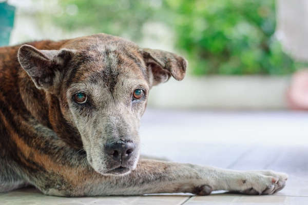
#6: Nuclear sclerosis in dogs
If you have a middle-aged or senior dog, and you catch a glimpse of his or her eyes from the side, you may notice that the lens looks cloudy and blue. This is due to a condition called nuclear sclerosis in dogs (lenticular sclerosis), which tends to affect dogs who are six to eight years of age. It is painless, doesn’t significantly affect a dog’s vision, and occurs as part of the normal aging process of the eye.
Cause of nuclear sclerosis in dogs
To understand the cause, it’s helpful to know that a dog’s lens is made up of numerous fibers, and over time, new fibers develop over the older ones. Some veterinary researchers feel that nuclear sclerosis is caused by the compression of these older fibers. This would explain why the cloudiness starts in the center of the lens and can work its way outward.
Symptoms of nuclear sclerosis in dogs
It is often easiest to see this blue-grey cloudiness of your dog’s lens when viewing the eye from the side rather than the front. It almost looks like your dog’s pupil is cloudy, but really, what you are seeing is the lens.
Because the lens is located behind the iris, you won’t see any cloudiness on the cornea or in the anterior chamber unless a different disease process is also present. Also, nuclear sclerosis is not a painful condition, so there shouldn’t be any squinting, inflammation, or other clinical symptoms.
Nuclear sclerosis makes the lens a little less flexible after a while, but dogs can still see reasonably well. To put it simply, if your dog were an avid reader, it would just mean he or she would need some reading glasses!
Diagnosis of nuclear sclerosis in dogs
Your vet can often easily diagnose nuclear sclerosis using a simple ophthalmoscope (a small magnifier with a light on it). If he or she can see through the lens to the back of the dog’s eye and there are no distinct areas of opacity, the dog has nuclear sclerosis. But if any opacities are present, the dog may have cataracts, which is the condition nuclear sclerosis most often gets mistaken for.
Treatment for nuclear sclerosis in dogs
There is no treatment for nuclear sclerosis. But since it is unlikely to affect a dog’s overall quality of life, treatment wouldn’t be necessary even if an effective one existed.
#7: Cataracts in dogs
Unlike nuclear sclerosis, cataracts of the lens can affect vision. A cataract in dogs is a white, cloudy spot (opacity) that forms in the center of the lens and can work its way outward. The cloudiness of a cataract is much denser than that seen with nuclear sclerosis. Thus, it does interfere with the way light travels through the eye.
To conceptualize this, think about the difference between looking through two windows. One window has a piece of plastic wrap over it (lenticular sclerosis). The other window has a piece of thick construction paper over it (cataracts). As you can imagine, the difference is significant.
Causes of cataracts in dogs
Primary cataracts can occur simply due to old age. Over time, proteins that come from lens fibers can accumulate like a growing snowball in the center of the lens. However, primary cataracts are also hereditary and may more commonly affect the following breeds, even in their younger years:
- Cocker Spaniels
- Boston Terriers
- Labrador Retrievers
- Australian Shepherds
Cataracts may also occur secondary to trauma or another illness. In dogs with diabetes mellitus, the excess glucose in their bloodstream can travel to the eye. Enzymes in the lens convert glucose to sorbitol, a very large molecule that attracts a lot of water. As a result of the excess water, the lens becomes cloudy. Interestingly, diabetic dogs can develop cataracts very quickly, literally within 24-48 hours of their diabetes diagnosis. So, if your newly diabetic dog woke up with cloudy eyes, cataracts might be the culprit.
Symptoms of cataracts in dogs
Besides observing a white opacity just beyond the iris (the cataract in the lens), you may notice other signs of cataract formation, such as redness and squinting. Additionally, your dog may suddenly have more difficulty seeing, especially at night. In cases where the cataract developed seemingly overnight, uveitis (and the related signs) may show up at the same time. This happens because the immune system releases inflammatory mediators to attack the cataract as if it were a foreign invader.
Diagnosis of cataracts
Based on the clinical signs and the appearance of the lens, your vet can determine whether your dog has a cataract (or cataracts). He or she might use an ophthalmoscope to determine how big the cataract is and its stage of development. For example, cataracts in the incipient stage take up less than 15% of the lens, while mature cataracts encompass greater than 75% of the lens.
Since diabetes mellitus can be related to cataract formation, your vet might also recommend blood and urine testing. Or he or she may recommend additional eye tests or other diagnostic tests, depending on the suspected underlying cause.
Treatment for cataracts in dogs
The treatment of choice for cataracts is a specialty surgery performed by veterinary ophthalmologists. It is called phacoemulsification and involves removing the old lens and replacing it with an artificial lens. Dogs with cataracts are considered good candidates for cataract surgery as long as any other illnesses (such as diabetes mellitus) are under control, and the dog’s retina is still functional.
It is very important to monitor your dog’s eyes carefully after cataract surgery and follow the veterinary ophthalmologist’s instructions carefully. If your dog develops cloudy eyes after cataract surgery or any other post-op complications, it’s essential to contact the surgeon immediately.
If your dog is not a good surgical candidate or if the surgery is cost-prohibitive, topical anti-inflammatory medicines may provide some relief from any discomfort that your dog may experience. At this point, there are no medications or supplements that can definitively treat cataracts in dogs.
Cloudy eyes in dogs FAQs
All this information certainly raises some questions. While your vet will be able to answer specific questions about your dog, we can discuss some of the more common questions that arise when it comes to cloudy eyes in dogs.
Do cloudy eyes in dogs mean blindness?
Cloudy eyes can have a range of impacts on vision. Some conditions do not affect vision, some cause partial or temporary vision loss, and some can cause blindness. Sometimes the same condition can have a variable impact on vision, depending on the severity.
For example, immature cataracts may cause partial vision loss, whereas mature cataracts result in complete blindness. Cataracts can be progressive, but do not always advance to cause full blindness. Both immature and mature cataracts cause cloudy eyes.
Why does my dog have a blue haze on his eyes?
When I think of blue cloudiness in dogs’ eyes, I think of corneal edema. As we’ve already discussed, corneal edema has several causes, so a vet will have to perform a physical examination and some diagnostic tests to figure out the cause and best course of treatment.
Interestingly, there is a disease often called “blue eye” that we very rarely see in dogs now—Canine Adenovirus Type 1. Adenovirus is one cause of hepatitis in dogs, but it also causes corneal edema.
Although this virus used to be quite common, numbers have decreased drastically due to vaccination! Adenovirus is usually included in the Distemper/Parvo combination vaccine for dogs. This is another reason it’s essential to follow your vet’s vaccination recommendations for all your pets!
When should I worry about my dog’s eyes?
I try not to be alarmist, but eyes can go very bad very quickly. I generally consider any sudden change in the eye, whether that is cloudiness, discharge, squinting, pawing/rubbing at the eye, etc., an emergency.
First of all, eye injuries hurt! Even sometimes putting in my contacts askew is agonizing. If for no other reason, I recommend urgent veterinary care to treat your dog’s pain.
Secondly, we often have the best chance to treat an eye condition quickly and completely if we address it early. The longer a condition is allowed to progress, the more difficult it will be to treat. In some cases, delayed treatment may even mean permanent vision loss. So, if you notice new cloudiness or any other change in the appearance of your dog’s eyes, the time to worry is now.
What should I do about cloudy eyes in dogs?
Understandably, if you suddenly notice that your dog’s eyes are cloudy, you want to do something to help your pup. Unfortunately, though, the reasons for cloudy eyes in dogs can be complicated. And treating the condition isn’t as simple as reaching for a bandage from your first aid kit.
Since that is the case, the best thing you can do is to call your veterinary clinic right away to schedule an exam for your dog. That way, you know that your dog is getting the help he or she needs. Eyes can worsen quickly, so eye problems (especially those that come up suddenly, are painful, or involve red and/or cloudy eyes) often require urgent veterinary assistance.
Treatment for cloudy eyes in dogs
As you may imagine, with such a wide variety of causes, treatment for cloudy eyes in dogs can vary. Some treatment is fairly simple and straightforward, whereas other conditions require more intensive care. Treatments for cloudy eyes in dogs depend on the underlying issue, and may include:
- Corneal ulcers
- Topical antibiotic ointment or drops
- Pain medication
- Serum drops
- Surgical corneal graft
- Corneal dystrophy
- Topical saline drops
- DSEK surgery
- Keratoconjunctivitis sicca (KCS)
- Topical eye lubricants
- Topical immunosuppressants
- Parotid duct transposition surgery
- Glaucoma
- Topical medications
- Corrective surgery
- Enucleation
- Uveitis
- Topical steroid or anti-inflammatory medications
- Systemic pain medications
- Topical or systemic antibiotics
- Nuclear sclerosis
- No treatment necessary or available
- Cataracts
- Phacoemulsification surgery
- Topical anti-inflammatory medications (for secondary problems, will not directly treat cataracts)
As you can see, some causes of cloudy eyes in dogs can be medically managed, and others are more effectively treated surgically. Some do not require treatment, and others do not currently have effective treatments available.
If a dog has a painful or otherwise uncomfortable condition that can’t be successfully treated, enucleation—surgical removal of the eye(s)—may be an option. Although removing the eyes is everyone’s last choice, the quality of life for a blind dog is often better than suffering from a chronic, painful condition. There are many ways you can care for a blind dog that will allow him or her to live a long, happy life.
Are there any home remedies for cloudy eyes in dogs?
While I do think that eye problems are best addressed by your veterinarian, there are a few “do’s and don’ts” of home treatment for cloudy eyes in dogs that you might find helpful.
DON’T use leftover eye ointment from another pet or problem.
Even if you do have an eye ointment in your doggie medicine cabinet, generally it isn’t a good idea to apply it to your dog’s eye without veterinary guidance. For example, some medications have a steroid as an active ingredient. If you apply that ointment to an eye with a corneal ulcer, it will delay healing or possibly make the ulcer worse.
DO stick to “safe” eye medications to care for your dog’s cloudy eye at home.
In general, most simple eye lubricants and eye irrigation solutions are safe for use in dogs’ eyes. Lubricants can soothe a dry eye and are usually not a problem when an infection or any other disease is present. Irrigation or saline solutions are useful for cleaning cloudy dog eyes after an injury or if there is a lot of discharge visible.
However, ensure that the lubrication or flushing products you use do not have “red eye reducer” or other added ingredients. These extra active ingredients may not be safe for your dog. If in doubt, consult your veterinarian before using a product.
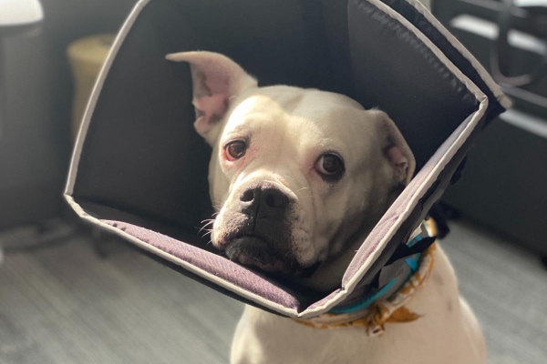
DO use an E-collar to protect the eye.
If your dog is rubbing at the eye or you are worried that there is a deep ulcer, put an Elizabethan collar on your dog to keep him or her from further traumatizing the eye.
DO NOT be fooled by products that claim to cure cloudy eyes.
Over-the-counter supplements or eye drops that promise to make your dog’s eyes brighter and healthier are available, but you must take these bold statements with a grain of salt. There are currently no supplements available that can fix cloudy eyes in dogs.
Some products like Ocu-GLO contain a mix of omega-3s and vitamins, which have anti-inflammatory and antioxidant properties, respectively. These are safe and can help with overall eye health. But ultimately, the way to treat a dog’s cloudy eye will depend on the disorder that is present.
Prevention of cloudy eyes in dogs
In some cases, cloudy eyes cannot be prevented. They may be caused by age-related changes or may be acute or unnoticeable until cloudiness sets in.
If your dog has underlying conditions that may predispose them to developing cloudy eyes, work with your vet to try to minimize the chance of ocular damage. For example, keeping diabetic dogs well-regulated decreases the chance of them developing cataracts. Treating dogs with KCS decreases the likelihood that they will develop permanent corneal damage.
Remember that if you have concerns about your dog’s eyes, seek veterinary care sooner rather than later.
Should you go to the vet if your dog has cloudy eyes?
As you have learned, young, middle-aged, and old dogs can have cloudy eyes for a variety of reasons. We have discussed the top ones here, but there are other conditions that could be the culprit, too.
If you notice your dog has cloudy eyes, it is best to seek immediate medical attention. Doing so can help avoid permanent damage and vision loss. Plus, since some eye conditions can be quite painful, getting to the vet ASAP means your dog can get some much-needed relief. Together, you and your vet can develop a plan to get your dog with cloudy eyes feeling, and hopefully seeing, better.
Why were your dog’s eyes cloudy?
Please comment below.
This post was originally published on October 5, 2022 and updated on September 29, 2025.

My dog is just turned 8 months old yesterday. I noticed he was squinting his left eye on Monday this week and overnight it has turned white (tuesday morning). I took him to the vet 2 days after (Thursday this week) and it was unclear what the cause of his cloudy/white eye was. The doctor prescribed tobramycin eyedrops and tolfenol and they did a CBC test and found out that he has a very little infection. The medication was recommended to use for 7-10 days only and if the won’t improve we need to go back to the vet for a blood chem but unfortunately I cannot afford it ATM because I just started college and I’m in my first year. I paid 1,600 for his CBC, consultation, and injection to reduce his fever and the 2 medication given. What should I do if it doesn’t get any better?
Dear Rhea,
I am sorry your puppy is having this issue with his eye. I understand how hard it can be to do what is best when finances limit the options. Unfortunately, there is really nothing I can recommend that can be done without returning to the vet. Eyes are very delicate and if things are not improving and the appropriate care is not administered in a timely manner, it could cause permanent changes or loss of eyesight. Make sure your vet is aware of your financial concerns and be honest about your wishes and limitations. They may be able to work with you or offer resources for support. Hoping all is well and praying for a full recovery for your sweet pup.
My dog is almost 4 years old and suddenly has a cloudy eye what seems like overnight. He’s a mix breed and has one white/blue eye and one brown. It is the brown eye that’s affected. He doesn’t have any other symptoms of anything that I can tell. I’ve been reading about the different causes of overnight cataract and he is fine otherwise than the sudden cloudy eye. His eating habits are the same, his energy, water intake, weight, everything. What can I do, other than going to the vet? Can I check blood glucose with a meter for humans or put some kind of eye lubricant in if it’s dry?
Hi Jennifer,
I understand your concern for your pup with this sudden change to his eye. I know you are trying to avoid a vet visit, but that’s exactly what we are here for. Human glucose meters can give false readings since they are calibrated differently and there is no way to know if the eye is dry without doing a tear test at the vet’s office. Eyes are delicate and if issues are not addressed quickly it can lead to permanent changes and possibly loss of vision. I strongly urge you to get your pup’s eye evaluated as soon as possible. Hoping for an easy solution and a positive outcome for your boy.
Hi, my dog is only 10 months old and has this large cloud in the middle of her pupil. The color of her eye had also changed. She is a Siberian Husky with ice blue eyes, but one of them was a dingey blue with some yellowing and the white part of the eye was red. I took her to the vet, and she was diagnosed with Conjunctivitis and then referred to an ophthalmologist for the cloud in the pupil. We are on the last day of the ten-day treatment for the conjunctivitis and now the other eye has developed a moderate-sized cloud, OVERNIGHT! I’m really concerned. We were finishing the meds before her Ophthalmologist appointment, and now I’m more concerned. Any idea what would cause a dog so young to develop these clouds so fast???
Hi Beth,
I understand your concern for your young puppy with these sudden changes to her eyes. I am not sure what the cause could be but think it would be best to get to the ophthalmologist as soon as possible. How are things today? Were you able to get a definitive diagnosis? Feel free to leave an update and let us know how things are going. Praying for healing and a favorable outcome for your sweet girl.
My 9 year, old black Chihuahua has whitish cloudiness covering both eyes. The vet says nothing can be done because it’s glaucoma. But every once in a while, the cloudiness in her eyes completely disappears for hours at a time, but never in both eyes at the same time. The vet says it’s not possible, but I have seen it and have photos. Is it possible she has been misdiagnosed?
Hi Janette,
I understand your concern for your pup with this strange cloudiness to her eyes that seems to randomly come and go. I am not sure I have seen anything quite like you describe but that’s not to say it isn’t possible. I do think it might be worth getting a second opinion. You may want to consider asking for a referral to a veterinary ophthalmologist. Hoping for clear answers and praying for many happy days ahead for your sweet girl.
my 12yrold terrier lost her left eye to glaucoma 7yrs ago the right is white but they’ve always been white since birth and today there’s green pus coming from left eye she seems in pain what’s going on with that eye ? I Don’t have any money for the vet and I’m so scared for my girl what can I do for her ? I can’t see this poor girl suffering
Dear Shelly,
I am so sorry your senior girl is experiencing this worrisome issue with her eye. It definitely sounds like an infection is present and this needs to be addressed right away. I understand finances are a concern, and I wish I had a good solution. Unfortunately, I am not aware of a way to resolve this from home without medical attention. I recommend you reach out to your local humane society or any rescue organizations in your area. They may have funds for these types of situations and might be able to help. Praying you can get your girl the care she needs. Bless you both. ♥
Hi Dr. Julie.
I have a Yorkie, he is 1.6 years old. He got a cloudy eyes in both eyes. It just happened in 2 hours. 1 night he got out and played outside while we are asleep, after 2 hours I woke up and seen his both eyes are cloudy, and reddish on the sides.
I think it got infected with the cement and mud he played on.
I have consulted to a vet, they told me he have a glaucoma. I’m afraid they got it wrong, since we’re living on an island and we only have 1 vet, who are not well-trained.
Hi Nori,
I am sorry your little pup is experiencing these worrisome eye issues. There are many possible causes, but without being able to personally examine your boy, I can’t make specific conclusions or recommendations. Did your vet prescribe some eye drops? How are things today? Hoping you have seen some improvement, and your boy has been able to maintain his vision. Please leave us an update if you have a chance. Praying for healing and many happy days ahead.
My dog started to have cloudy eyes , took him to the vet I had noticed he started bumping into things at night at first and then I took him to the vet right after. They told me there was nothing he could do that my dog will go blind. I had mentioned he had diabetes but all he said was it’s bc of the diabetes that 99 percent of dogs go blind their end of their first year of being diagnosed with diabetes but my dog is currently 5 months into being diagnosed. That’s it was a cataract that he got due to his diabetes. He didn’t run no test on him on his eyes just sorted looked at him and that’s it. Now my dog can’t see anymore but he doesn’t bump into things as much as before. I heard there was a surgery to remove their lens into an artificial lens. I want to know how late and how do you know when you dog is fully blind
Hi Daniela,
I am sorry your dog has developed cataracts, and his vision has been impaired. What your vet said about dogs with diabetes developing cataracts is true. While there are specialty centers that offer cataract surgery, not every dog is a good candidate, and this treatment can be very expensive. If you want more information about surgery, it would be best to ask your vet for a referral to a veterinary ophthalmologist. They will be able to go over all the details and offer advanced testing to determine if your pup still has any degree of vision left. Hoping you can get the answers you need to feel comfortable with how to proceed. Wishing you and your sweet boy all the best.
Hi Dr. Julie,
My 6.3 kg poodle was diagnosed with PRA, based on my statement that she lost night vision when she was 2. Now she is 7.7. A year ago a cataract started to form and we were managing it with eye drops. However, in March she got bitten/stung/ by a yellow jack(yellow bee), and cataract fully developed in 2 weeks, in both eyes. Based on ERG, a doctor from UK mentioned that the equipment is missing some data and readings are not clear to proof it is a PRA, but definitely there is something wrong with the retina (it was an online consultation), but still a PRA is not to be proofed by the ERG. A local ophthalmologist is suggesting a surgery. I have read that after a bee stung it is possible to have an eye nerve inflammation (in humans), which cannot be seen sometimes. I wonder if this is a possibility, based on the toxic/poison from the yellow bee. I am just looking for any suggestions/advise you can give me in that case. She was seeing big objects as cats before that, and now she cannot see, since the cataract fully developed. Thank you!!!
Hi Dilyana,
I am sorry your little pup has been through so much over the past few months. Honestly, ophthalmology is not my expertise, and I would rather defer to the specialist for how to proceed. If you have not discussed the bee venom theory with the ophthalmologist, then I would encourage you to do so. Don’t hesitate to speak up and advocate for your girl. Hoping you can get the answers you need to feel comfortable with the treatment plan moving forward. Wishing you the best of luck and praying for a positive outcome.
hii
I have a 2 nd half year old doberman today while checking up on her I have seen grey cloudiness on her left eye. I don’t think he has any problem in vision she is not lethargic or bumping into anything . she is acting normal but the cloudiness is there. what would be the reason.
Hi Priyanka,
I understand your concern for your young Doberman with this sudden change to her eye. As the article states, there are many reasons an eye can look cloudy and the only way to narrow down a cause is to do some investigation. Without being able to examine your pup myself, I can’t make specific conclusions. No matter what the cause, eye issues need to be addressed very quickly as they can progress at a rapid rate and have the potential to cause loss of sight. Please contact your vet as soon as possible so they can get your girl the medical attention she needs. Best of luck to you both!
hi
I have a 15 months Shih tzu, since Friday he has closed his eye and while checking up on him I have seen blue grey cloudiness. I have seen vet and he has given some medicine including blood based serum. also I don’t think he has any problem in vision bcoz he is not lethargic or bumping into furniture. he is acting normal but the cloudiness is there. what would be the reason.
Hi Shaun,
I am sorry your senior guy is having issues with his eye. Unfortunately, without examining him myself, it is hard to make specific conclusions. When there has been an injury to the cornea or an ulcer present, the cornea can sometimes take on extra fluid (corneal edema) and this can make it look cloudy. If this is the case for your pup, then it may go away with more time or it may be a sign that things are not completely healed. I would encourage you to discuss this with your vet and see if they think a recheck exam is needed. Hoping all is well and praying for a full recovery for your sweet boy.
yes, he was in fight with cat and she injured him. when I am puting eye drops to his eye, I have seen a blood clot near cornea and his eye is also red. can you suggest whether this damage is permanent or temporary.
Hi Shaun,
I understand your concern for your pup and this injury he has sustained to his eye. There really is no way to make an assessment on if the eye issue will be able to completely resolve without personally examining your boy. Do you have a recheck scheduled with your vet for some time in the near future? If not, I strongly encourage you to reach out and offer them an update. Eye problems need to be monitored closely as things can progress rapidly and lead to loss of vision in the eye if not properly addressed. Wishing your sweet boy all the best and praying for complete healing.
My dog is a three year old white shepherd. Her left eye suddenly becam light sensitive 2 days ago. It is cloudy and red. She won’t let me touch it. Opens wide and she’s fine otherwise. I a, snowed in from a Colorado blizzard and won’t be out for about a week. I have some amoxicillin from a vet and given her some.. any advice? Thanks.
Hi Denise,
I am so sorry you are in this tricky situation with your Shepherd. From what you describe I am suspicious that she may have scratched her cornea or somehow injured her eye. While the oral antibiotics may help some, your girl really needs to have her eye evaluated, and it will probably require medicated eye drops. I know you can’t help the weather, and this is out of your control. It just has me very concerned because eye issues can progress quickly and lead to the loss of vision and possibly the loss of the eye itself. Try to keep your girl from rubbing her eye! It is imperative to seek medical attention as soon as you can get her to the vet or find someone to make a house call. Praying for healing and wishing you both the best. ♥
My 14 yr old ShihTzu X Maltese has been pretty much problem free, other than the seemingly harmless few bumps found on parts of his body. On Saturday he wok up with his right eye closed and covered with a lot of discharge. He kept it closed most of the day and I checked on him throughout the day. On Sunday morning I opened his eyelid and found the eye was cloudier and the whiteness also covered a much larger area comparing to the other eye. I could not get him to see a vet on Sunday so I cleaned it with a wet napkin. Both two days he was quieter than usual and eating about half of his typical meal. By Sunday afternoon I noticed him walking slower and bumped into a table, I am now convinced his right one can no longer see. On Monday I took him to a vet and was told the possibility of glaucoma, cataract, and dry eye. I was recommended to give him eye drops. The vet was speaking English as a 2nd language so I could only understand about 30% of his words. Does my dog have any chance at regaining his vision at this point? Would the same thing happen to his left eye without any warning like the right eye? What should I do going forward to prevent this issue to his left eye? Thanks so much.
Hi Shera,
I am so sorry your boy is dealing with this painful eye issue. While there are many possible causes, there are tests that can be done to help narrow down the problem. Does your vet have a way to check eye pressures? Or can they stain the eye to make sure there is no scratch or ulcer present on the cornea? Without examining your pup myself, it is hard to make specific conclusions. There are eye conditions that can cause sudden blindness, but from what you describe I would not think that is the case for your boy. It may be necessary to get a second opinion or even ask for a referral to a veterinary ophthalmologist. Hoping you can get the answers you need and praying for healing and relief for your senior guy.
my dog is 11 years old and has suddenly woken up today with cloudy eyes since he was a pup he always had a hue of blue over them but today they are really cloudy like grey, i have him booked in tomorrow but just wondering if any one had this, he is well in himself drinking eating urinating, even being playful, he will let me look in his eyes but unsure if hes in any pain, as he does squint when im looking at him, the outsides look a little bloodshot too, he was in the bin the other night, im not sure what he had eaten but wondering if its a possibility something has triggered this.
Hi Rosie,
I am sorry your senior guy is having issues with his eyes. I think you made a wise choice in taking him to your vet. There are just too many possible causes to mention them all in this reply. But eye problems can progress quickly and may lead to blindness, so it is always important to have them checked right away. How are things today? Was your vet able to make a diagnosis? Hoping all is well and praying your boy is able to make a full recovery.
Hi Dr Buzbys,
I have one Shiba Inu and she is 2years and 8months old. From last week I have notice that her right side eyes is becoming small and little cloudy thr. But when we go for walk and play she open her both eyes wide.
I took her to the vet and they did fluorescein stain tests and vet didn’t saw anything but Vet and we all can see little cloudy in her eyes. She gave us eye drop 3 times a day for one week bcuz vet said it might be infection from something. Am so worrying and what you think about her eye.
Hi Tenzin,
It sounds like you are headed in the right direction. I am glad the fluorescein stain was negative, and your vet is not worried about a corneal ulcer or scratch. Without examining your pup, myself, I can’t make specific conclusions. I hope the antibiotic drops will help resolve the problem and your girl will start feeling better quickly. Keep in touch with your vet and let them know if you don’t see the improvement that was expected. Best wishes!
My dog just had a lensectomy; her eye has a dent where the incision was made and it looks like a cloud is seeping from the indent. I am very worried she has a visit tomorrow however I am scared the doctor might suggest enucleation or brush off my concern I sent photos of this and she said it looked fine. My dog has an obvious white scar where the incision was made and is not acting lethargic.
Hi Kasey,
I understand your concern for your dog, and I am glad you had a visit already scheduled with the vet. How did things go? Were there any issues found during the recheck? Feel free to leave an update if you have a chance. Hoping your sweet girl is doing well and was able to make a full recovery.
Hi this is Curtis, Carson is our 12 + years old Labradoodle. He just started taking your pills. He had a bowel movement in the house in the middle of the night. He has had a problem of late doing that until we had to stop giving him gabapentin, which helped a lot in that area. He still has these accidents from time to time but rarely.
Will the new pills affect that in any way?
We know as adults that when you start a new med it can affect your system for a an awhile before it clears up. Carson is over 100 lbs and we gave him 3 of the pills spread out through the day.
What do you think?
Thanks so much, we are hoping for the best for Carson’s pain and mood
Hi Curtis,
Thank you for reaching out about Carson. I am glad you have decided to try Encore Mobility to help with pain control in your senior guy. I generally do not see any GI side effects in patients taking Encore Mobility but am not sure how it will affect Carson’s current defecation issues. Has your vet found a cause for the fecal incontinence? Most dogs of advanced age will start to show some symptoms of canine cognitive dysfunction/dementia and incontinence (urine or feces) can be one of the early signs. I have had many clients with dogs diagnosed with CCD say that Encore Mobility helped to lessen the severity of symptoms. Maybe it will help Carson in this area as well? Please feel free to leave an update and let me know how things are going. Hoping you see the positive results you need to improve your sweet boy’s quality of life.
I have a mongrel dog he’s almost 1 year old. he came in yesterday with a closed eye, now today he has a cloudy eye, a small amount of discharge. he doesn’t cry when I touch it. could this be due to a scratch or injury as opposed to a disease/condition? also. if so, will it heal?
Hi Heather,
I understand your concern for your young dog with these eye issues that have emerged. A closed eye is usually a sign of pain. All the symptoms you mention have me very worried that your pup may have endured an eye injury that needs to be addressed right away. Eye problems can progress very quickly and end up in the loss of the eye. Also, eye injuries can be extremely painful causing suffering if not treated properly. Please contact your vet as soon as possible. Hoping the eye can heal and your sweet boy can get some relief!
I adopted my cocker spaniel Roxy three years ago when she was eleven. Her groomer noticed left eye was looking somewhat reddish and “off”.Groomer works at our vet clinic and said to get her checked ASAP.. Over the course of several weeks IOP spiked up and down. We opted to have her seen at vet school for ophthalmic exam. Diagnosed with primary glaucoma. She lost the sight in left eye, pressure spiked to 85, so we elected a chemical ablation.. It was the right decision for us. Three years later still sees out of right eye, very thankful. She also has dry eye. Yes, there are drops, but that’s not a problem. Thankful groomer spotted problem and primary. Veterinarian keeps track between visits to opthamologist. Great article!
Hi JoEllen,
Thank you so much for sharing your experience with our readers. This is a perfect example of how everyone can come together to work as a team for a beloved dog’s best interest. Isn’t it great to know you have such amazing people backing you up every step of the way? I am glad Roxy is doing so well and her daily treatments are manageable. Best wishes and bless you both!