When it comes to a corneal ulcer, dogs and humans alike would agree that it is painful. Not only that, but left untreated, a corneal ulcer could cause significant damage to a dog’s eye. Integrative veterinarian Dr. Julie Buzby explains why corneal ulcers occur, how to recognize one, and what your vet will do to help your dog feel better. Plus, she gives some instructions on applying eye medication.
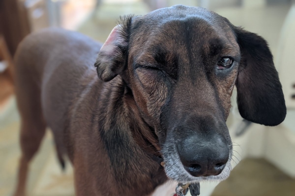
A dog with a painful, cloudy, or squinting eye is always an emergency in my book. Granted eye issues are not life-threatening, but they can be sight-threatening. Plus, eye pain is miserable. If you’ve ever gotten an eyelash or a grain of sand stuck in your eye—or worse, had a corneal ulcer—you know what I’m talking about.
Recently, my own dog, Jake, had a corneal ulcer. The poor guy was miserable when it happened. But thankfully with some quick treatment, he was back to himself in no time. This experience reminded me of how important it is that dog parents have the scoop on ulcers. And that’s where this article comes in.
- What is a corneal ulcer?
- Canine corneal anatomy
- What are the types of corneal ulcers in dogs?
- What are the causes of a corneal ulcer?
- What are the symptoms of a corneal ulcer in dogs?
- How is a corneal ulcer diagnosed?
- How are corneal ulcers in dogs treated?
- An "eye" on the future
- Has your dog ever had a corneal ulcer?
What is a corneal ulcer?
To start off, let’s break down the term “corneal ulcer” a bit. “Ulcer” is a medical term used to describe an erosion at the surface of an organ or tissue that is accompanied with necrosis (i.e. cell death) of the surrounding tissue. Then “corneal” refers to the cornea, which is the clear portion at the front of the eyeball. So a corneal ulcer would be an erosion of the cornea.
Before we go any further, let’s take a look at the anatomy of the cornea. This information will be handy when discussing the different types of ulcers.
Canine corneal anatomy
The cornea functions as the “windshield” of the eye. It protects all the other important ocular structures that enable normal vision. The cornea itself is typically 0.62mm thick, or about half the thickness of a dime, and consists of four layers that all serve an independent purpose.
- Corneal epithelium—the outermost layer of the cornea which is coated by the tear film. It is made up of stratified, nonkeratinized squamous cells in a layer that is 5 to 7 cells thick. These cells form tight junctions with each other and are water repelling, or “hydrophobic” to keep the tear film from seeping in. The cells constantly turn over. This means the body can generate a whole new epithelial surface in 7 to 10 days.
- Corneal stroma—a finely woven network of tightly organized parallel collagen fibers. The stroma supports corneal development and healing. This layer is water absorbing, or “hydrophilic.”
- Descemet’s membrane—a very thin tissue layer, only 7 micrometers thick, that separates the stroma from the endothelium. This tissue layer is quite elastic and will curl inwards if disrupted.
- Endothelium—a single cell layer of hexagonal cells that regulates the water content of the cornea to help keep it clear.
The cornea also contains nerves. In fact, it is one of the locations in the body with the highest density of nerve fibers. So, it is no wonder that damage to the cornea—like a corneal ulcer— is so painful.
What are the types of corneal ulcers in dogs?
Just as there are different parts of the cornea, abnormalities in each of these parts result in different types of ulcers. Each ulcer type has a potentially different treatment plan and outlook.
Superficial corneal ulcer
These ulcers only affect the epithelium and have a high success rate of healing rapidly with appropriate treatment. The corneal epithelium is the fastest-healing tissue in the body. This means most superficial ulcers will heal in five to seven days as long as there is no concurrent infection, and your dog isn’t rubbing or scratching at the eye as it heals.
Indolent corneal ulcer
Also known as persistent corneal erosions or Boxer ulcers (because Boxers are predisposed to them), this type of ulcer has a ring of loosely adhered epithelium around the border of the ulcer and has been present for at least two weeks without healing.
Unfortunately, these ulcers are a bit more tricky to treat. This is because while the stroma is intact, the loose cells at the edges make it difficult for the body to get new epithelial cells to stick to, and heal, the defect. As a result, these ulcers often persist for weeks or even months.
Deep corneal ulcer (i.e. stromal ulcer)
Damage that is deeper than the epithelium will penetrate the water-absorbing layer of the corneal stroma, creating a deep or stromal ulcer. These ulcers can be more complicated to manage.
Melting corneal ulcers in dogs
Sometimes neutrophils (i.e. immune cells), microorganisms, or corneal epithelial cells produce enzymes that may inappropriately break down the collagen within the stroma. Since the cornea appears to be melting away, these are called melting corneal ulcers. Unfortunately, melting ulcers can quickly become severe and require rapid and aggressive treatment to try to save the eye.
Descemetocele
This is the term for ulcers that penetrate to Descemet’s membrane. Since Descemet’s membrane is one cell layer thick, it is very easy for the eye to rupture. When the ulcer perforates, aqueous humor (i.e. the fluid in the front chamber of the eye) leaks out. This hole will become blocked from the inside by a fibrin clot or the iris. Descemetoceles, especially those that have perforated, require emergency surgical intervention to prevent the dog from losing the eye entirely.
Simple vs complicated ulcers
Additionally, vets may use these descriptors for corneal ulcers in dogs:
- Simple (i.e. uncomplicated)—only involves epithelium and does not progress into the stroma or persist for longer than seven days.
- Complicated—involves the stroma (i.e. deep ulcers and descemetoceles), does not heal within seven days (i.e. indolent ulcers), becomes infected, and/or starts melting.
What are the causes of a corneal ulcer?
Now that you have an understanding of some of the terminology surrounding corneal ulcers, it is time to address what makes a corneal ulcer occur in the first place. Corneal ulcers are particularly common in dogs. This may be due to their large eyes and tendency to traumatize their eyes during normal activities or play. However, this is not the only way your dog can get an ulcer. Other causes of corneal ulceration include:
- Foreign bodies—Getting a plant awn, piece of grass, splinter, etc. in the eye can cause corneal injury and ulceration.
- Endocrine disease—Systemic disease such as diabetes, hypothyroidism in dogs, and Cushing’s disease in dogs can all predispose your dog to recurrent corneal ulcers.
- Anatomic disorders—Congenital abnormalities such as eyelids that roll inward (i.e. entropion) or abnormally growing lashes (i.e. distichia) can cause constant rubbing on the corneal surface, resulting in ulcers. Having eyelids that roll out (i.e. ectropion) also can make ulcers more likely.
- Poor quality or decreased tear production—Keratoconjunctivitis sicca (KCS) (i.e. dry eye in dogs) leaves the corneas unprotected by sufficient tear film and more prone to scarring, ulceration, and infection.
- Oversized eyes—Some breeds such as Pugs and Shih Tzus have eyelids that cannot close completely over their corneas to protect and re-wet them. This results in dry and damaged corneas that are more likely to develop a corneal ulcer.
- Epithelial dystrophy—This inherited condition of some dog breeds causes them to be born with a weakened corneal structure, making them prone to corneal ulcers.
What are the symptoms of a corneal ulcer in dogs?
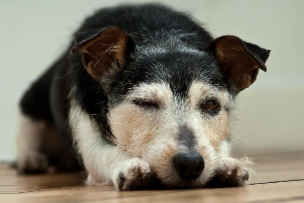
As mentioned previously, the cornea is richly innervated, which means that corneal damage is quite painful. You may notice your dog squinting or holding their eyelid closed (i.e. blepharospasm). Some dogs may rub or paw at the eye, which runs the risk of making the ulcer even worse. Additionally, you may see other signs your dog is in pain. The eye may be watering or you might see some mucoid discharge.
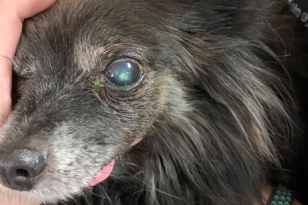
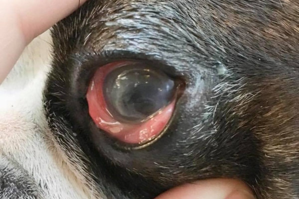
Dogs with a corneal ulcer also may have an area of bluish or whitish cloudiness to the cornea (i.e. corneal edema). This is not the same as age-related changes to the lens (i.e. nuclear sclerosis in dogs) or opacity of the lens (i.e. cataracts in dogs). The cloudiness is on the surface of the eye with a corneal ulcer and within the eye (in the lens to be exact) with nuclear sclerosis or cataracts.
If the ulcer has been there a bit, you may also notice little red lines running across the cornea or forming a red border around the edges of the cornea. These are new blood vessels, which are attempting to come into the cornea and help it heal. Normally, the cornea does not have blood vessels.
If you notice any of these symptoms, please call your vet immediately. When it comes to eye problems, time is of the essence. Please do not take a “wait and see” approach.
How is a corneal ulcer diagnosed?
The symptoms listed above indicate that there is an eye issue. But they are not specific to corneal ulceration in dogs. Your vet will need to perform some diagnostic tests to figure out if your dog has a corneal ulcer or some other eye problem like glaucoma in dogs. During the appointment, your vet may take an approach that looks something like this:
Observe from a distance
Don’t be surprised if your vet wants to watch your dog from across the room at first. He or she is looking for any difference in symmetry to the face and eye sockets. Additionally, the vet will be assessing how your dog holds the eyelids when the face is unrestrained and whether or not the eyes appear to be visual.
Examine the eyelids
Constant rubbing from abnormal eyelids or eyelash growth can cause corneal ulcers in dogs. So the vet will want to pay attention to the orientation of the eyelids and location of the eyelashes. Abnormalities of those structures may require surgical correction as well as treatment of the ulcer. Otherwise, your dog is likely to continue to suffer from corneal ulceration.
Neurologic examination
The vet will also want to assess your dog’s neurologic system as it relates to the eyes. The goal is to be able to find the answer to these three questions:
- Does your dog respond to visual cues normally in both eyes?
- Can he or she still blink appropriately?
- Do the dog’s pupils respond to light stimulus? Or are they stuck in either a dilated or constricted position?
Schirmer Tear Test
The vet will insert a special folded piece of paper under the lids of both eyes. This is a good way to measure tear production over one minute. Low tear production indicates KCS, which can predispose dogs to corneal ulcer.
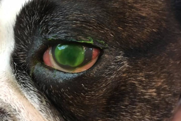
Fluorescein stain
This is arguably the most important diagnostics tool when examining a corneal ulcer. The vet will apply a drop of painless, special stain to the corneal surface. Then he or she will immediately flush the eye with eye wash. Next, the vet will turn the lights in the exam room off and use a specially filtered light to examine the cornea for residual stain.
Because it is hydrophobic (i.e. water repelling), a healthy and intact cornea WILL NOT retain the water-based stain. However, with damaged corneal epithelium, the exposed hydrophilic (i.e. water loving) layer of the stroma WILL take up the stain. This stain will appear as a bright green dot or scratch under the light.
Finally, if your dog has a descemetocele, the stain distribution will look like a donut. In other words, the stroma will be green because it takes up stain. But there will be an unstained crater in the middle, which is the hydrophobic Descemet’s membrane.
How are corneal ulcers in dogs treated?
Once your vet has diagnosed your dog with a corneal ulcer, he or she will discuss the treatment plan with you. Management of your dog’s ulcer will depend on its type, cause, and severity.
Pain control
First of all, pain management is key. Ulcers are extremely painful, and relieving this discomfort with systemic and/or topical pain control is part of any good treatment plan. Dogs may benefit from non-steroidal anti-inflammatory drugs (NSAIDs), gabapentin for dogs, tramadol for dogs, or other pain medications.
Treatment for superficial corneal ulcers
This type of ulcer has a good success rate with proper management as long as it remains uncomplicated. Your vet will prescribe topical antibiotics to address infection. He or she may also potentially use a medication called atropine to prevent eye spasms and provide additional pain relief.
Your dog will blink away the medications very rapidly. So it is important to make a plan that allows for you to apply the drops or ointment multiple times per day. The good news, though, is that a superficial ulcer usually heals within five to seven days.
Treatment for deep/stromal corneal ulcers, descemetoceles and melting ulcers in dogs
These types of corneal ulcers require more extensive treatment due to their severity.
Some may require a stronger antibiotic, both oral and topical antibiotics, and/or more frequent administration of antibiotics. Topical autogenous serum (i.e. the fluid part of your dog’s blood) or other medications may help decrease the breakdown of corneal collagen that occurs in melting ulcers.
When surgery may be necessary
If the dog’s ulcer continues to worsen despite aggressive treatment or is deeper than 50% of the stroma, surgery may be the best option. A board certified veterinary ophthalmologist can use a flap of the dog’s conjunctiva (i.e. pinkish tissue on the surface of the eyeball) or a biosynthetic graft to cover the ulcer. This supports the cornea, protects the ulcer, and delivers a direct blood supply to promote healing.
Treatment for indolent corneal ulcers in dogs
These ulcers also are tricky to treat. The normal healing process of the epithelial tissue is being undermined since the cells can’t stick to the corneal stroma. In these cases, your vet may recommend surgical intervention to remove, or “debride,” the non-adherent tissue with a cotton swab. The vet may then perform a grid/punctate keratotomy.
Alternatively, the vet may decide to do a diamond burr debridement and keratotomy. Both the grid/punctate and the diamond burr keratotomy procedures alter the surface of the cornea in that region to make it easier for epithelial cells to adhere and complete the healing process. Many ulcers heal within a month or so of treatment but some take longer or require additional procedures.
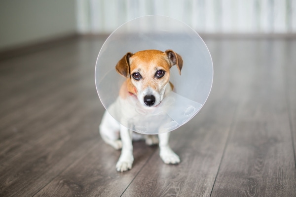
The importance of E-collars
In virtually all cases of ulcer healing, your vet will provide you with an Elizabethan collar (E-collar) for your dog to wear. Corneal tissue is extremely fragile, especially when it is healing over damaged tissue. For this reason, it is critical to protect the eye from further trauma. Otherwise, you run the risk of having the ulcer worsen or the eye rupture. So please always have your dog wear the E- collar during the healing process!
Address the underlying cause
In addition to managing the ulcer, it is important to think about why the dog had the ulcer in the first place. If it was trauma-related, then this was probably a stroke of bad luck. But if there is an underlying cause like metabolic disease, dry eye, etc. then it is a good idea to try to get those issues under control too, and hopefully, prevent ulcers in the future.
Work closely with your vet
There are no appropriate “home remedies” for the treatment of corneal ulcers. Ulcers are painful, and with delayed or incorrect treatment, can deteriorate quickly to the point that your dog’s eye may not be salvageable. So if you suspect your dog may have a corneal ulcer, you should not try to treat it yourself. Instead seek the guidance of a veterinary professional.
Since ulcers can change rapidly, keep a close watch on your dog’s eye. If your dog suddenly seems more painful or the eye looks worse (i.e. increased cloudiness or redness, abnormal discharge, etc.), you should contact your veterinarian immediately.
Additionally, your vet will probably want to recheck your dog’s eye periodically until the ulcer heals. Plan to bring your dog to those appointments even if it seems like the eye is doing better.
Get comfortable with applying eye medication
Many of my clients feel intimidated by the idea of applying eye medications multiple times per day. I get it. It can seem daunting at first. Hopefully, your veterinary team along with the following instructions can help boost your confidence.
First off, it’s a good idea to ask your veterinarian or the veterinary nurse to demonstrate the proper technique during the appointment. Then you can do a practice administration in front of them so you feel more comfortable with it.
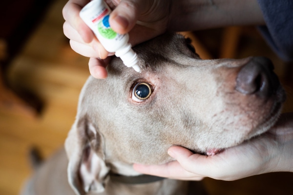
For the first few medication administrations, it may be helpful for you to muzzle your dog or have someone help you hold your dog’s head. Even the most well behaved and loving dog can snap when painful or scared. Corneal ulcers are definitely painful at first, and having drops put in the eye is unsettling for some dogs.
Steps for administering eye medications to your dog’s eye
To help you with the process, I have included the following steps for you to follow when applying eye drops or medications to your dog’s eyes:
- Hold the ointment or dropper tube using your dominant thumb and pointer finger and rest your hand on top of your dog’s head while you stand behind them. Use your other hand to point their face up towards the ceiling while supporting under their jaw.
- Use the thumb or a finger of your non-dominant hand (the one not holding the tube) to pull down the lower eyelid. Be careful that you do not put too much pressure on the eyeball itself when you do this. The injured cornea is fragile and the ulcer could worsen or the eye could rupture with excessive pressure.
- Squeeze the ointment or drops onto or inside the edge of the lower eyelid. Never touch the tube of medication to the eyeball or lids themselves as that can contaminate the medication.
- Release your dog’s head and offer lots of praise and a treat. Usually even the most difficult patient quickly learns to tolerate eye medications as their level of pain decreases with treatment.
- If you have multiple medications to administer, ask your veterinarian in what order to use them and how long between each administration. That way you’re not just washing one medication out with the other.
For more help, especially if your dog is fearful or nervous about eye medications, check out Veterinary Partner’s fantastic explanation of applying eye drops with low stress handling.
An “eye” on the future
I know that when you walk out of the vet clinic with a bag full of eye medications and a dog who is running his or her E-collar into everything, it can seem a bit overwhelming. But the good news is that superficial eye ulcers generally heal quite quickly, especially if you catch them early. Other types of ulcers do present a bigger challenge, but with appropriate treatment and patience, you and your dog will come out on the other side before you know it.
Has your dog ever had a corneal ulcer?
Please comment below.

We had our golden pup recently spayed about 16 days ago. Her spay healed great but the initial day of surgery I noticed she wouldn’t open her left eye, I called and told my vet and agreed it may be due to her still being out of it and on heavy meds and I would see if it improved overnight. The eye did seem to improve, she never really acted painful/pawed at her eye/face. Two days later the symptoms resumed and she wouldn’t open her eye again so we took her to the vet and they found the “ pretty large” superficial eye ulcer. They gave us antibiotics ointment and we had a recheck in a week. At the recheck the ulcer seemed unchanged and they just increased the frequency of the eye antibiotics. My concern is that there was no change, and now a day after our recheck my dog’s eye almost seems worse now after they looked at it. I’m thinking of getting a second opinion but the factor is my current vet did own up to this being caused from them as a complication of the spay surgery and they are paying for everything, but now I don’t feel like they’re doing what should be done/ what they are recommending is not giving any improvement. My husband and I do not leave our pup alone at all( we thank goodness have that luxury with our work schedules) and watch her every move to try to make sure she doesn’t harm the healing process- the poor pup has been in an ecollar since her spay and confined to very limited activity. I just don’t understand how I feel we’re doing everything we were told to do and there’s no improvement and now almost seems worse. Am I just overthinking this, or should I go somewhere else for treatment? Also a side note is our vet said they would send us to an eye doc if there was no improvement after another week of the current line of treatment but I feel like if it’s not improving then maybe we should just go to an eye doc
Dear Erin,
I am sorry your pup is dealing with this worrisome eye injury. What a blessing your vet was honest about this accident and is willing to do whatever it takes to get your girl on the road to recovery. Eyes can be tricky and, despite our best efforts, sometimes things just don’t heal as we would hope. It sounds like they are following the standard of care for this type of situation and have “plan B: already in place (a trip to the ophthalmologist) should things not start to improve. If you would like to move forward with seeing the specialist don’t hesitate to make your request known. Hoping all is well and your girl’s eye is completely restored. Feel free to leave an update if you have a chance.
I’m desperate for help. My 6 year old Frenchie has recurring corneal ulcers.
Hes had allergies his whole life as is typical with these dogs. He’s on Apoquel, first 5.4 mg a day and now 16 split into two doses, 8mg am and pm.
He still loves to rub his face on things (really just the couch or my bed) when he’s happy and playful. He injures his eye at least every other month.
His vet says he doesn’t have eyelash or eyelid issues. The abrasions respond to Tobramycin, however he does have residual cloudy spots in both eyes from having these so often.
We discussed debridement however my vet didn’t recommend it for now since he does still rub his face.
I’m at a loss!! He is not exhibiting any other allergy symptoms on the apoquel. Vet finds no skin fold issues at all, nothing else on his face or body that indicate an issue. He doesn’t scratch himself or lick when he’s on the medication.
His groomer also finds him to be very healthy and no signs of skin issues.
He has no vomiting, diarrhea, nothing at all. No indication of any other illness.
He just won’t stop rubbing his face in things when he’s being goofy!
I work from home, so he’s near me all day. I do stop the behavior most of the time. But I’m exasperated for myself and sad for him. He has to wear an e collar so often, and that’s no way to live. What should i look into??
Hi Jodi,
I am sorry you are dealing with this frustrating issue with your Frenchie. From what you describe it does sound like it could be purely behavioral. Could you find something to lay over the couch and your bed during the day that would deter him from wanting to rub his face? I know some people use the plastic mats made to go on the floor under a desk chair to put on couches to prevent dogs from getting up on the furniture. I am not saying this is the right product for what you are facing, but just wondering if you could brainstorm a good idea. You may also want to talk with a boarded veterinary behaviorist as they can help offer training advice for behavioral modification. Your boy is lucky to have you advocating for his health and well-being. You are doing a great job. Wishing you both the best of luck and keep up the good work.
Thank you for your detailed article. Our 14y/o female terrier mix needs either a cornea graft or her eye removed. Unfortunately she had large piece of the cornea came off and her eye is close to rupturing. Our vets opinion is a cornea graft is a “heroic measure” and isn’t strongly advising us either way. Her other eye also has some of the same cornea ulcer that we are treating but currently is manageable. Do you have an opinion on either procedure?.
Hi Gabrielle,
I am sorry your pup is dealing with these severe eye issues. I have never performed a corneal graft and therefore don’t have much information on if this offers a better solution that removing the eye. I do know that I have had many patients that were missing an eye (either from enucleation surgery or injury) and they all seemed to feel great and live normal lives. I am not sure there is a wrong answer here and you would be doing your pup justice with either option you choose. Hoping you can find the answers you need to feel confident moving forward with treatment. Best wishes and feel free to leave an update as things progress.
Hi guys
My dog has 2 really bad eye ulcers .
He’s been on isathal and remand and stromease and optixcare. It been 3 weeks and he still has them maybe a little better but not much. We have seen the vet lots and costing so much money.
Any advice please 🙏
Thank you ☺️
Hi Karen,
I am sorry your pup is dealing with ulcers that just won’t heal. It sounds like it may be time to ask about a referral to an ophthalmology specialist. Sometimes surgical intervention is needed in these more complicated cases. Praying for answers and a clear path forward.
Hello! Our little Luna , Morty,Poodle mix, was just diagnosed this condition after bringing her home from the groomer, I called the groomer the next morning and told them my Luna was not able to open her left eye and they quickly arranged for me to take her to an Emergency Vet where they told me she had the ulcer, they gave her medication and pain meds to take home and hopefully she will be fine in about two weeks
Hi Victor,
I am so sorry Luna is dealing with this painful eye condition. What a blessing your groomer was willing to step up and arrange medical care for her! Hoping her recovery is quick and praying she will be back to her normal self very soon.
my shih-tzu was diagnosed with an eye ulcer,eye drops for 4 x’s daily for 7 days & liquid meds in food or orally for 5 days,goes back to the vet to re-check in 1week.
Hi Charlotte,
I am sorry your pup is dealing with a painful eye ulcer. Hoping you will start to see improvement very soon and praying for complete healing.
Hello,
Our senior Dachshund woke up a week ago Friday with her eye closed we got her into our vet, the stain test showed a horizontal superficial ulceration below her pupil. She was given the serum and an antibiotic drop. Giving her the serum drops every 2-3 hrs, antibiotic drops every 12 the ulceration appeared almost healed by Monday but the lower lid still red swollen our vets took her back to check under 3rd eyelid there was a tiny superficial ulcer there. She has more in pain since that was done two days later we took back again ulcer doesn’t appear to be worse but the tear test in her other eye was just on the edge so vet had us use artificial tears over the weekend and reduce the serum to 2x daily. She seems more painful but her eye itself doesn’t appear worse. She has liver issues so giving Gabapentin is risky, should she still be this painful 10 days out?
Hi Brenda,
I am sorry your Dachshund is dealing with this painful eye injury. It is a bit worrisome that it has been treated for 10 days and still is painful and does not seem to be healing appropriately. It may be time to think about a referral to a veterinary ophthalmologist. They have tons of experience dealing with more complicated cases and might have some treatment options that are not available in general practice. Hoping things will start to resolve and praying for a positive outcome.
Many thanks for your helpful article. Our 18 year old chihuahua’s left eye has literally just collapsed inward and we have an appointment with our veterinary ophthalmologist first thing in the morning. She is crying and appears to be in pain. She has other issues such as abdominal pain (from medications) and diarrhea. My wife has been taking her to various vets regularly and this new eye problem severely compounds matters. I’m overseas on a work assignment and am thinking of just coming home to help. Given our dog’s age and other ills we’re not sure if she can withstand surgery. We are distraught and not sure how to proceed. Your thoughts would be very much appreciated.
Hi Dean,
I am so sorry your senior girl is in this difficult situation, and you are out of the country. Despite your dog’s other issues, surgery to remove the eye may be the only option. How did the ophthalmologist appointment go? Hoping they had some good advice and a clear path forward. Feel free to leave an update if you have a chance.
Good article, you are the only one whom states details on what a ruptured ulcer may look like. We have an appointment with an eye doctor tomorrow for our 18 year old Papillon who we suspect has a ruptured ulcer after about 3-4 weeks. Initially it was on the road to healing beautifully but had a set back last week when he bumped his eye (has bad cataracts and almost blind) then it deteriorated. We have only had him about 4 months as he was my dad’s dog and he passed away recently. We are learning a lot about caring for a blind/ poor visioned dog. Hopefully there is something we can do for him, but it might be possible he will have to have it removed.
Dear Carlene,
I am sorry your senior guy is having so many eye issues. Bless you for taking care of your dad’s beloved pup. Praying you received good news from the ophthalmologist. Hoping you were able to avoid surgery and there were options for medical management. Feel free to leave an update if you have a chance. Wishing you all the best. ♥
Two weeks ago my 15.5year old Shih Tzu had a descemetoceles which ruptured. It all seemingly happened overnight. We got him to an ophthalmologist asap – 36hrs – and he underwent a graft surgery, setting me back a cool ~$6k. He was very dysphoric and back legs were like noodles coming off the anesthesia. He is since back to his typical evening restlessness / begging for food and treats routine, with spurts of wobbly walking and legs slipping esp. on hard surfaces. Wearing the cone and antibiotics left him susceptible to ear infection which we’re managing on top of it all. He’s had two good rechecks, albeit it was a little more raised than ideal. Vet had us start amniotic drops to hopefully kick start the healing and flatten it out. After his 4th dose it seemed he was tearing more and possibly more raised. It seemed like one of the baby stitches may have dislodged. I could have gone in to see the on call / after hours vet, but i felt like that would be more traumatizing for my little guy and wouldn’t want to put him under again to fix the stitch. We’re stopping the amniotic drop for now and giving him some time to chill. I’m losing my mind, second guessing whether I made the wrong choice and should have removed it. He had just had a good senior wellness visit a month ago, kidney issue he’s had since a young age was well managed, and now it feels like everything is falling apart. He’s still a happy dude, tail wagging (less so on the Gabi) and strong appetite. I just bought the toe grips to see if that’d help with the steadiness at least. Any advice?
Hi Trish,
I am sorry your senior guy has had so many issues with his eye. I understand your concerns about the delayed healing. Unfortunately, since I haven’t personally examined your dog, it is hard to make specific recommendations. It sounds like you are doing everything you can to give your boy the best shot at a full recovery. I am hopeful ToeGrips will give your sweet pup some extra traction when he walks. Please let me know what you think about them! I hope you can find the answers you need to navigate this unknown path. Wishing you both the best and praying for complete resolution of this eye problem for your dog.
Thank you for the article. My dog has a deep cornea ulcer . We are treating it with medication three days out it seems to be getting better. Doing a follow up in seven days to see if it’s continuing to get better. If it is not getting better definitely will have surgery. ($3,800) Reading your article has helped me understand why, and how eye is i ulcer developed in my dogs eye.
Hi Tally,
Thank you for the kind words about the article. I am glad you found it informative and helpful. I am hopeful your dog’s eye will continue to heal and you will be able to avoid surgery. Praying for a speedy and complete recovery. Feel free to leave an update after the next checkup if you have a chance.