As a dog parent, a basic knowledge of dog leg anatomy could come in handy when talking to your vet or researching a new issue that popped up for your beloved canine companion. Integrative veterinarian Dr. Julie Buzby discusses the anatomy of a dog’s front and hind legs in easy-to-understand terms. And she also gives an overview of some of the more common conditions that affect different areas of a dog’s leg.
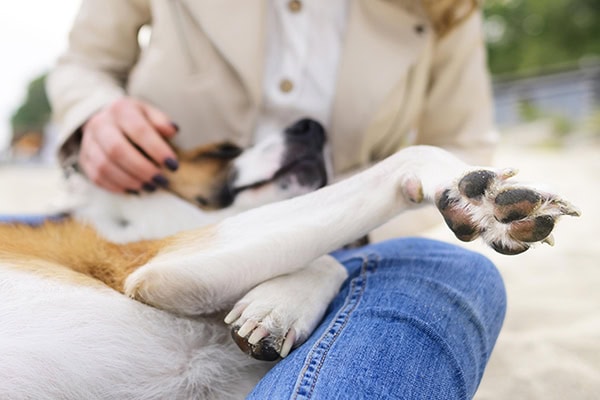
While human and dog leg anatomy have a lot of similarities, at times it may still be difficult to find the right word for the different parts of your dog’s legs. The hips and shoulders translate pretty well between dogs and humans. But as you move down the leg it can get a bit more confusing.
For example, let’s take this one-question quiz:
Is your dog’s knee:
A) The joint in the middle of the front leg?
B) The joint semi-hidden within the thigh of the back leg?
Answer: B The knee is in the back leg. What you see in the front is really the dog’s carpus (wrist).
If you chose answer “A”, don’t fret! By the end of this article, it will be easier to name the different parts of your dog’s legs!
Why not call your dog over and use him or her as a living anatomy model as we start this journey into dog leg anatomy together.
Dog rear leg anatomy
Since more of the common orthopedic issues in dogs affect the back legs, they are a good place to start. While it isn’t a perfect comparison, it can sometimes help to picture a dog’s back leg as being similar to yours if you were standing on your tip toes and folded over at the hips.
Dog hip anatomy
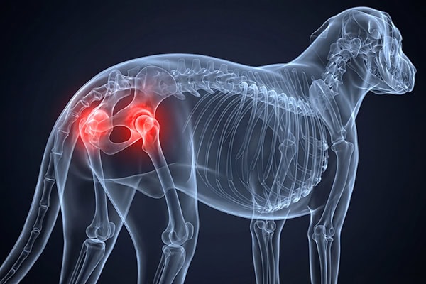
If you place your hand on top of your dog’s pelvis and slide it toward one of the rear legs, the first joint you will come to is the hip, or coxofemoral, joint.
Two of the bones that make up the hip joint are part of the pelvis—specifically the ilium and pubis. They meet to form a semi-circle indent called the acetabulum. This is the “socket” part of the hip joint.
The rounded top of the femur, which is the longest bone in the body and extends down into your dog’s thigh, provides the “ball” part of the hip joint. The head of the femur sits within the acetabulum. Several strong ligaments, muscles, and tendons help hold the femur in place and provide stability for the hip joint.
Problems associated with a dog’s hip
The two main issues dogs have with their hips are:
- Hip dysplasia: In hip dysplasia in dogs, the hip joint doesn’t form properly while the dog is growing, causing the head of the femur and the acetabulum not to fit together well. This instability of the hip joint can lead to pain and arthritis over time.
- Hip dislocation: Due to trauma such as a fall, being hit by a car, or landing wrong, the head of the femur may come out of the acetabulum. Having hip dysplasia may also make a dog hip dislocation more likely to occur.
Dog knee anatomy
Continuing on down the hind leg, the bottom of the femur (thigh bone) and the top of the tibia (shin bone) meet to form the knee, or stifle, joint. Between the femur and tibia lies a cartilage cushion called the meniscus that helps distribute compressive forces.
Plus, like humans, dogs have a patella (knee cap), which sits within the patellar tendon in a groove at the bottom the femur. The patellar tendon, which comes off of the quadriceps muscles, attaches to the front of the top of the tibia.
Two sets of ligaments help stabilize the knee joint. The medial and lateral collateral ligaments run down the inside and outside of the knee and connect the femur to the tibia. They are responsible for preventing those bones from moving side to side relative to each other.
The other set of knee stability ligaments are the caudal and cranial cruciate ligaments. They form an “X” shape (hence cruciate in the name) and are found within the knee joint. These ligaments have the important job of keeping the tibia from sliding backward or forward under the femur.
Knee problems in dogs
There are three main problems that may affect a dog’s knee:
- Cranial cruciate ligament (CCL) rupture: Degeneration of the CCL over time leading to weakening of the fibers, or much less commonly, sudden trauma to a healthy ligament, can lead to a partially or completely torn ACL in dogs. Some dogs will also suffer a meniscal tear.
- Patellar luxation: Due to improper knee anatomy such as a shallow patellar groove, or sometimes trauma, the patella may slip out of the patellar groove. There are different grades of luxating patella in dogs depending on how severe it is.
- Osteosarcoma: Some of the most common locations for osteosarcoma in dogs, a type of bone cancer, are the bottom of the femur or the top of the tibia. (The others are the bottom of the radius and top of the humerus in the front leg.)
Dog hock anatomy
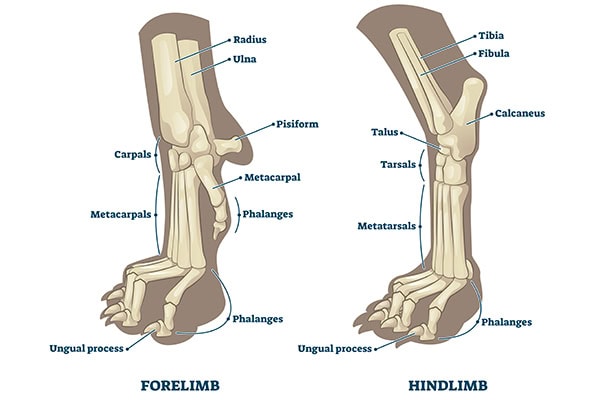
The bottom of the tibia and fibula (thin bone that runs parallel to the tibia), the tarsal bones, and the tops of the metatarsal bones (analogous to the bones in your feet) form the tarsus or hock. This is the equivalent of a dog’s ankle.
Several ligaments help support the hock. And the Achilles tendon attaches to the calcaneus (pointy bone that is like your heel bone.)
Hock injuries
While problems with the hock are less common than those of the knee or hip in dogs, they do occur. Examples include:
- Achilles tendon (common calcaneal tendon) rupture: Cuts or other trauma to the Achilles tendon or chronic degeneration may cause it to rupture. Affected dogs may stand with the hock dropped or touching the ground.
- Hock fractures: Racing Greyhounds especially, but other dogs too, can fracture bones in their hock.
- Ligament injuries: Injuries to the supporting ligaments of the hock such as sprains or ruptures may cause instability and pain.
Rear paw anatomy
To form the foot, the four long thin metatarsal bones meet up with the toe bones (phalanges). There are three sets of toe bones that make up each toe, and the toenail covers the tip of the third phalanx (P3). Other important parts of dog paw anatomy are the paw pads and the tendons and ligaments.
Most dogs don’t have rear dewclaws (the claw located midway between the foot and hock). However, there are some breeds like Great Pyrenees who should normally have double dewclaws on both rear feet.
Paw problems
Some potential paw problems in dogs include:
- Broken toes or metatarsals: Trauma such as getting stepped on or getting a toe caught or twisted may cause fractured metatarsals or broken toes in dogs.
- Paw pad injuries: Burns, cuts, flaps, and other paw pad injuries in dogs are possible.
- Broken nails: Dogs who rip a nail off or split a nail are often limping and licking the area due to the pain.
Dog front leg anatomy
Now it’s time to look at front leg anatomy. If you lean forward and pictured putting your weight on the base of your fingers and tucking your elbow in at your side, this is a rough representation of your dog’s front leg anatomy.
Dog shoulder anatomy
Starting from the top again, the first joint you come to on the front leg is the shoulder. It is made up of two bones—the scapula (shoulder blade) and the humerus (upper arm bone). The rounded head of the humerus sits in a “cup” at the bottom of the scapula. This makes the shoulder joint a ball and socket joint like the hip.
Shoulder issues
Some potential shoulder problems in dogs include:
- Osteochondritis dissecans (OCD): OCD occurs when a cartilage flap develops on the joint surface, leading to inflammation and pain. It is most common in rapidly-growing large breed puppies and can also occur in the elbow, knee, or hock.
- Supraspinatus tendinopathy or biceps tenosynovitis: Dogs may develop inflammation, tearing, or scaring of the tendons of the biceps or supraspinatus (large muscle on the portion of the shoulder blade closer to the head). Supraspinatus tendinopathy or bicep tenosynovitis tend to be more common in working or sporting dogs, or other active dogs.
Dog elbow anatomy
The humerus continues until it meets the radius and ulna (forearm bones) to form a dog’s elbow. This joint functions like a hinge and is one of the more common locations for front leg orthopedic issues.
Elbow dysplasia
Most of the issues in the elbow can get grouped under the umbrella of elbow dysplasia. This includes:
- Osteochondritis dissecans (OCD): Just like in the shoulder, dogs can develop a cartilage flap in the elbow joint (usually on the humerus) that leads to pain and inflammation.
- Fragmented medial coronoid process (FCP): The coronoid process, a tiny bony protrusion on the ulna that makes up part of the lower portion of the elbow joint, breaks off. The end result is pain and instability.
- Ununited anconeal process (UAP): Sometimes the anconeal process, a small bony wedge at the head of the ulna that forms the back of the elbow joint, does not properly fuse to the rest of the ulna. This creates a painful bone fragment within the joint and makes the joint less stable.
Dog carpus anatomy
Moving down the dog’s forearm, the next joint is the carpus (wrist). Like the hock, the carpus contains rows of small carpal bones. Above the carpal bones are the bottom of the radius and ulna. And below them are the tops of the metacarpals (long thin bones like the bones in your hand). These bones are stabilized by connective tissue.
Carpal problems
Carpal hyperextension in dogs is the main carpus-specific condition to be aware of. It occurs when trauma, degeneration, abnormal development, or other conditions weaken the supporting structures, allowing the carpus to sink down closer to the ground.
Front paw anatomy
The anatomy of the front paw is essentially the same as that of the back paw. Front dewclaws are much more common than rear dewclaws, although some breeders may have them removed shortly after birth. The same problems that affect the rear paws can also affect the front paws.
Some diseases may impact multiple joints
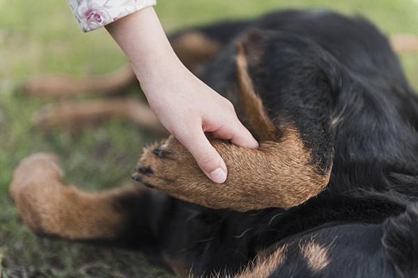
While we have discussed some of the leg injuries or conditions that affect specific joints, it is important to remember that there are also conditions that can impact multiple joints.
By far, the most common one is arthritis. Osteoarthritis in dogs is a progressive inflammatory condition involving degeneration of the joints, pain, and reduced mobility. Many of the conditions already discussed, such as hip dysplasia, patellar luxation, and ACL tears, can contribute to a dog developing arthritis. So can obesity and a variety of other factors.
Additionally, some infectious diseases, especially tick borne diseases in dogs like Lyme disease, can cause inflammation in multiple joints. So can immune-mediated polyarthritis (IMPA in dogs), which occurs when the immune system attacks the joints. In both cases, the dog may seem lame on one leg one day, and another leg a different day. This is called shifting leg lameness.
If your are concerned, make an appointment with your vet
Hopefully, your expanded grasp of dog leg anatomy will help you better advocate for your dog and communicate with your vet. If you are concerned because your dog is limping, has a swollen joint, doesn’t seem to be moving around as well, or is otherwise acting abnormal, don’t hesitate to reach out to your vet. The sooner you address the problem, the better!
What questions about dog leg anatomy do you have?
Please comment below.